Download
1 / 1
10 likes | 200 Views
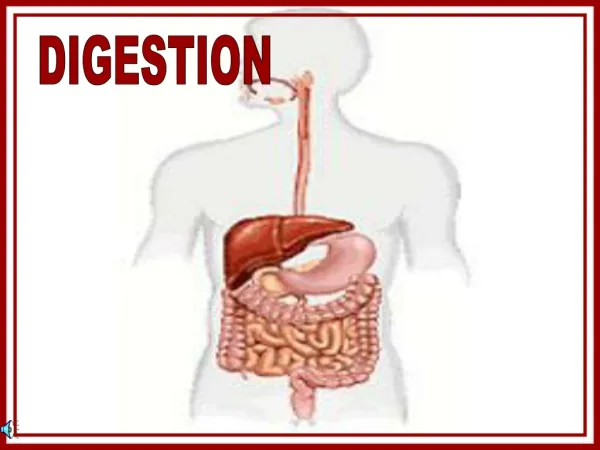
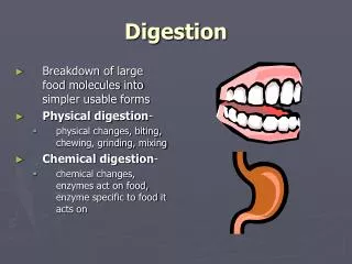
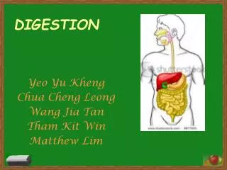
Organs 1. Alimentary canal a. Function i. Digests ii. Absorbs b. Organs i. Mouth ii. Pharynx iii. Esophagus iv. Stomach v. Small intestine vi. Large intestine. Large Intestine. Absorption. Small Intestine. Functional Anatomy.

E N D
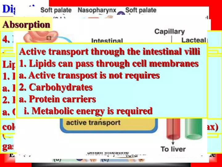
Organs 1. Alimentary canal a. Function i. Digests ii. Absorbs b. Organs i. Mouth ii. Pharynx iii. Esophagus iv. Stomach v. Small intestine vi. Large intestine Large Intestine Absorption Small Intestine Functional Anatomy Histology of the Alimentary Canal Digestive Process Stomach Overview Chemical Digestion Digestion 4. Intestinal phase: Excitatory phase: Duodenum releases a gastrin-like substance that promotes additional activity of gastric glands. Inhibitory phase: Enterogastric reflex inhibits vagal nuclei, inhibits local reflexes, activates sympathetic fibers that tighten pyloric sphincter & causes the release of enterogastrones. 5. Enterogastrone are secretin, cholecystokinin (CCK), vasoactive intestinal peptide (VIP) & gastric inhibitory peptide (GIP) Accessory digestive a. Organs i. Teeth ii. Tongue iii. Gallbladder b. Glands i. Salivary ii. Liver iii. Pancreas Essential activities 1. Ingestion a. Taking food into digestive system via the mouth 2. Propulsion a. Movement of food through the digestive tract b. Swallowing i. Voluntary c. Peristalsis i. Involuntary 3. Mechanical digestion a. Chewing b. Mixing i. Saliva c. Churning d. Segmentation i. Local constriction of the small intestine 4. Chemical digestion a. Mouth through small intestine Proteins 1. Begins in stomach a. Pepsinogen to pepsin 2. Continues in small intestine a. Trypsin and chymotrypsin i. Pancreatic enzymes b. Carboxypepsidase i. Pancreatic enzymes Four basic layers 1. Mucosa: Innermost layer functions in secretion, absorption and protection. It consists of 3 sub-layers: lining epithelium, lamina propria & muscularis mucosae. Epithelium: Simple columnar with goblet cells. Lamina propria: Loose areolar connective tissue with capillary beds & lymph nodules. Muscularis mucosae: Thin layer of smooth muscle 2. Submucosa: Composed of dense CT (blood vessels & lymphatic vessels) & elastic fibers 3. Muscularis externa: Functions in segmentation & peristalsis. It consists of inner circular and outer longitudinal layer of smooth muscle. Sphincters are thickened areas of smooth muscle 4. Serosa: Areolar connective tissue covered with mesothelium (single layer of squamous epithelium) Regulation of gastric secretion 1. Three phases: Cephalic, gastric & intestinal 2. Cephalic phase: Occurs prior to food entering stomach (brain response to food) via hypothalamic stimulation of the vagus nerve (parasympathetic enteric ganglionic neurons stimulate the stomach glands) Salivary glands 1. Function of saliva: Cleanses mouth, dissolves tastants, moistens & initiates chemical breakdown 2. Types: Extrinsic (3 pairs: parotid, subman-dibular & sublingual) & intrinsic (buccal glands) 3. Secretory cells: Serous cells (watery secretion with enzymes) & mucous cells (viscous) 3. Secretory cells of gastric glands: Mucous neck cells (produce acidic mucus), parietal cells (secrete HCl and intrinsic factor), chief (zymogenic) cells (produce pepsinogen which is converted initially by HCl into pepsin which will catalyze conversion thereafter) & enteroendocrine cells (produce hormones that regulate digestive function) Gross anatomy 1. Four regions: Cardiac (region where food enters), fundus, body & pyloric. Terminates at the pylorus & connects with small intestine via pyloric sphincter. 2. Greater curvature (lateral convex surface) 3. Lesser curvature (medial concave surface) Active transport through the intestinal villi 1. Lipids can pass through cell membranes a. Active transpost is not requires 2. Carbohydrates a. Protein carriers i. Metabolic energy is required Digestive processes occurring in stomach 1. Enzymatic digestion: Protein digestion is initiated in stomach (pepsin) 2. Lipid soluble substances can pass through stomach mucosa (alcohol and aspirin) 3. Production of intrinsic factor: Required for absorption of B12 Microscopic anatomy 1. Expanded surface area for absorption 2. Structural modifications: Circular folds (deep folds of the mucosa and submucosa), Villi (finger-like projections of the mucosa) & Microvilli or brush border (projections of plasma membranes) Digestive processes in mouth, pharynx & esophagus 1. Mastication: Mechanical breakdown by teeth and tongue 2. Deglutition: Complicated process of swallowing (two phases which involves 22 muscles) Esophagus 1. Laryngopharynx into esophagus 2. Pierces diaphragm & joins stomach via cardiac orifice gated by cardiac sphincter 3. Four layers: Mucosa, submucosa, muscularis (skeletal and smooth regionally distributed) & adventia (not serosa: entirely connective tissue) Background 1. Hydrolysis a. Catabolic process b. Large molecules into monomers c. Enzymes into lumen of alimentary canal i. Intrinsic and accessory glands Tongue 1. Bundles of skeletal muscle 2. Function a. Mix food with saliva i. Bolus b. Position bolus for swallowing Carbohydrates (starch) 1. Carbohydrates are broken down into glucose, fructose and galactose 2. Process a. Salivary amylase b. Pancreatic amylase Pancreas 1. Accessory digestive organ 2. Produces digestive enzymes: Exocrine product (pancreatic juice) 3. Acini: Secretory cells surrounding ducts 4. Composition of pancreatic juice: Proteases, amylase, lipases & nucleases 5. Absorption a. Movement of digested end products from lumen of the GI tract into blood and lymph 6. Defecation a. Elimination of undigested materials 5. Regulation of bile release: CCK from small intestine is released into blood in response to fatty chyme entering small intestine. CCK stimulates secretion of pancreatic juice & relaxes hepato-pancreatic sphincter (controls entry of pancreatic juice and bile entering duodenum) 4. Subdivisions: Cecum (1st segment: saclike), appendix (lymphatic dead end), colon (several regions: ascending, transverse, descending & sigmoid), rectum (contains rectal valves: internal transverse folds) & anal canal (two sphincters: internal involuntary and external voluntary) Gross anatomy 1. Ileocecal valve to the anus 2. Absorbs water from indigestible food residues 3. Unique features: Teniae coli (3 bands of longi-tudinal smooth muscle), haustra (pocketlike sacs) & epiploic appendages (fat-filled pouches) Teeth 1. Classification: Incisors, canines, premolars & molars 2. Dental formula: (2I, 1C, 2PM, 3M/2I, 1C, 2PM, 3M) X 2 = 32 Microscopic anatomy 1. Four tunics 2. Lining epithelium: Simple columnar (entirely goblet cells) with gastric pits (gastric glands that produce gastric juice) Lipids 1. Digestion occurs solely in small intestine a. Lipases from pancreas 2. Bile emulsifies fats so they are soluble a. Only increases area that enzymes can contact D. Liver and gallbladder 1. Accessory organs associated with small intestine 2. Liver has a role in digestion in addition to its other functions: Bile production and export (emulsification of fat) 3. Gastric phase: Local signaling within stomach due to the presence of food (distension, peptides or low acidity) lead to HCl release (gastrin is released in response to chemical stimuli & stimulates the release of HCl by parietal cells) Mouth (oral cavity or buccal cavity) 1. Boundaries: Lips, cheeks, tongue & palate 2. Palate: Hard palate (rigid & underlain by bone) & Soft palate (formed from muscle) 5. Digestive processes: No breakdown, just water and vitamin absorption & major function is the peristaltic movement of fecal material 6.Defecation: rectal walls stretch(defecation reflex) which is spinal-parasympathetic reflex (causes colon & rectum to contract & anal sphincters relax) Histology 1. Simple columnar epithelium 2. Submucosa includes lymphatic tissue: Peyer’s patches 3. Gallbladder: Stores bile 4. Bile: Only bile salts and phospolipids aid in digestion (emulsify fat) other components that do not contribute to digestion will not be discussed Stomach 1. Expansion of the GI tract 2. Sack for storage and chemical breakdown in which food is converted to chyme Regulation of pancreatic secretion: Local hormones (CCK & secretin) & parasympathetic nervous system Gross anatomy 1. Pyloric sphincter to ileocecal valve 2. Subdivisions: Duodenum, jejunum & ileum Nucleic acids 1. Pancreatic nucleosides Liver and gallbladder Pharynx: Oropharynx to Laryngopharynx Home Exit BASIM ZWAIN LECTURE NOTES