Download
1 / 40
420 likes | 805 Views
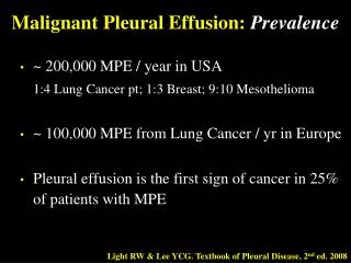
Diagnosis and Management of Malignant Pleural Effusion. 衛生署桃園醫院內科加護病房主任 莊子儀醫師 2006 年 7 月 20 日. Etiology of Malignant Effusion. Lung cancer: 37.5%, especially adenocarcinoma Breast cancer: 16.8% Lymphoma: 11.5%, most common in young adult. Etiology of Malignant Effusion.

E N D
Diagnosis and Management of Malignant Pleural Effusion 衛生署桃園醫院內科加護病房主任 莊子儀醫師 2006年7月20日
Etiology of Malignant Effusion • Lung cancer: 37.5%, especially adenocarcinoma • Breast cancer: 16.8% • Lymphoma: 11.5%, most common in young adult
Etiology of Malignant Effusion • Increasing production of effusion: • Increasing vascular permeability: invasion of pleural vessels by tumor, cytokines, injury, infection etc. • Increasing vascular hydrostatic gradient: decreased pleural pressure by atelectasis, increased venous pressure by SVC syndrome, decreased plasma osmotic pressure by hypoalbuminemia • Nonvascular entry by thoracic duct: chylothorax
Etiology of Malignant Effusion • Decreasing exit of effusion: • Increasing resistance to lymphatic flow: infiltration of parietal pleura or mediastinal lymph nodes by tumor seeding • Increasing gradient opposing lymphatic flow: decreased pleural pressure by atelectasis, increased venous pressure by SVC syndrome
Clinical Presentation • Dyspnea • Cough • Chest pain
Radiographic Evaluation • Chest X-ray
Chest X-ray • Amount of pleural effusion • More than 2/3 hemithorax or even entire hemithorax • 55% of large and massive effusions • Other causes: empyema and TB effusions • Cytology diagnosis of large and small effusions: no significant difference (63% vs. 53%)
Chest X-ray • Mediastinum position • Shift away from a large effusion • Midline mediastinum in large effusion: significant lung collapse, fixed mediastinum LAP • Shift toward a large effsuion: trapped lung due to main-stem bronchial obstruction
Radiographic Evaluation • Chest X-ray • Chest CT
Chest CT • Pleural surfaces, lung parenchyma, chest wall and mediastinum • Malignant pleural disease: pleural thickening (>1 cm), irregularity, nodules • Pleural thickening: also seen in empyema • Pleural nodules: only 17% in malignant effusions • Other features: lung mass, chest wall involvement, mediastinal LAP, hepatic metastases
Radiographic Evaluation • Chest X-ray • Chest CT • Chest echo
Chest Echo • Pleural surfaces, lung parenchyma, chest wall and pleural effusion • Pleural effusion: echo-free • Pleural thickening and nodules • Echo-guide thoracocentesis • Echo-guide pleural biopsy
Diagnosis • Pleural effusion • Cytology • Pathology
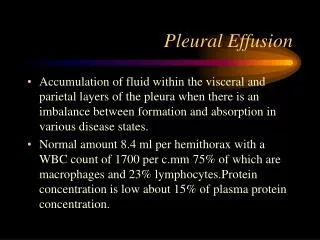
Pleural effusion • Grossly bloody: most common cause of bloody effusion • Serosanguineous effusion • Cell differentiation: lymphocytes predominant • Eosinophilia: can not exclude malignant effusion
Pleural effusion • Almost always exudate • Lactate dehydrogenase (LDH): increased cell turnover and lysis • Low glucose concentration and low pH level: possible shorter survival • pH < 7.20: easily failure of pleurodesis
Cytology • Adenocarcinoma: most likely to be positive • Low pH: greater tumor burden • Cytology diagnosis of large and small effusions: no significant difference (63% vs. 53%) • Body fluid + cell block
Pathology • Pleural biopsy • Closed needle biopsy • Cope needle • Abrams needle • Urocut needle
Diagnostic Procedures • Diagnostic thoracocentesis under echo-guide • Send pleural effusion for routine, BCS (LDH, protein, glucose), Gram/AFB stain, cytology, B/C, plus ABG (for pH) • Pleural biopsy under echo-guide • Send pleura for pathology and TB tissue/C • Therapeutic thoracocentesis under echo-guide • Send pleural effusion for body fluid + cell block
Primary Tumor (T) • T4: • A tumor of any size with invasion of the mediastinum, or involving heart, great vessels, trachea, esophagus, vertebral bodies, carina, • or with the presence of malignant pleural/pericardial effusion, • or exudative pleural effusion without evidence of obstructive pneumonitis, • or with satellite tumor within the lobe of primary tumor at chest CT
Management • Symptom-oriented management • Less than 1/3 hemithorax, C/T sensitive tumor • C/T at first, F/U regularly • Slowly recurring effusion, short life span • Repeated therapeutic thoracocentesis • More than 2/3 hemithorax, no airway obstruction • Pigtail insertion for pleurodesis within 24 hours
Management • Before pleurodesis • Daily drainage amount < 100-150 ml • Confirm with chest echo • Ability of lung re-expansion • Chemical pleurodesis • Mnocycline injection • Beta-iodine injection • OK-432 injection
Management • Pre-medication • 2% xylocaine 10ml in 50ml normal saline • Minocycline injection • After 15 minutes, 300mg Minocycline in 50ml normal saline • Clamp catheter/tube, change position 2-6 hours • Unclamp catheter/tube, low pressure suction
Management • Pre-medication • 2% xylocaine 10ml in 50ml normal saline • Beta-iodine injection • After 15 minutes, 10 ml beta-iodine in 40ml normal saline • Clamp catheter/tube, change position 2-6 hours • Unclamp catheter/tube, low pressure suction
Management • Indwelling catheter • Good outpatient situation • Good for trapped lung • Pigtail catheter with drainage bag • Chest tube with Heimlich valve
Complication • Re-expansion lung edema • Empyema • Restricted lung disease • Trapped lung
Prognosis • Medium survival • Lung cancer with malignant effusion: 3 months • Breast cancer with malignant effusion: 5 months • Mesothelioma with malignant effusion: 6 months • Lymphoma with malignant effusion: 9 months
Thank You for Attention • Reference: • Murray and Nadel’s Textbook of Respiratory Medicine, 4th edition, 2005 • Light and Lee’s Textbook of Pleural Disease, 1st edition, 2003 • Mathis and Lessnau’s Atlas of Chest Sonography, 1st edition, 2003