Download
1 / 25
250 likes | 385 Views
Retrieving and Viewing Protein Structures from the Protein Data Base. 7.88J Protein Folding Prof. David Gossard Room 3-336, x3-4465 gossard@mit.edu September 15, 2004. Protein Data Base. Established in 1971 Funded by NSF, DOE, NIH Operated by Rutgers, SDSC, NIST

E N D
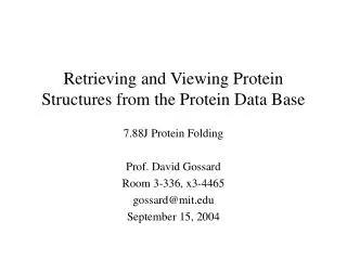
Retrieving and Viewing Protein Structures from the Protein Data Base 7.88J Protein Folding Prof. David Gossard Room 3-336, x3-4465 gossard@mit.edu September 15, 2004
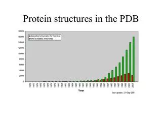
Protein Data Base • Established in 1971 • Funded by NSF, DOE, NIH • Operated by Rutgers, SDSC, NIST • Purpose: Make protein structure data available to the entire scientific community • In the beginning: “less than a dozen” protein structures • Currently has 27,112 protein structures • Growing at 20% per year • New structures 50 times larger than those in 1971 are commonplace
Why the “Knee in the Curve”? • Engineered bacteria as a source of proteins • Improved crystal-growing conditions • More intense sources of X-rays • Cryogenic treatment of crystals • Improved detectors & data collection • New method - NMR: • Accounts for 15% of new structures in PDB • Enables determination of structure of proteins in solution • “Protein Structures: From Famine to Feast”, Berman, et.al. • American Scientist v.90, p.350-359, July-August 2002
Why is the PDB Important? • Rapid, extensive access to new structure data • “Collective Leverage” for … • Understanding molecular machinery • Rational drug design • Engineering new molecules • Structural genomics • etc…
Not all Structures are Different • PDB Growth in “New Folds”
Structure vs Sequence • New protein sequences are being discovered much more quickly than new protein structures are being solved • Currently, known protein sequences vastly outnumber known protein structures • The “sequence-structure” gap continues to widen
Point of Information • Today’s material is: • a subset of the information available to you in online tutorials • presented to “get you started” quickly and to “shorten the learning curve” • not exhaustive or even sufficient => should be augmented by actually working through the online tutorials
PDB Website http://www.rcsb.org/pdb/ Enter what you know…
Query Result Browser Which one do I want? Let’s look at this one …
Structure Explorer Yep, that’s the right one… View it… Download it…
View Structure Static Images
Download/Display Display the file header… Download the file… (Select this file format)
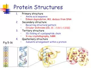
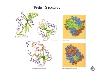
Visualizing Proteins • High complexity • Multiple levels of structure • Important properties are “distributed” throughout the 3D structure Branden & Tooze
Visualization Objectives • Structure • Backbone; secondary, tertiary & quaternary • Side chain groups • Hydrophobic, charged, polar, acidic/base, etc. • Cross-links • Hydrogen bonds, disulfide bonds • Surfaces • VanderWaals, solvent-accessible • Charge distributions, distances & angles, etc.
Display Conventions Wireframe Ribbon Spacefill Molecular Surface
History of Visualization of Macromolecules • http://www.umass.edu/microbio/rasmol/history.htm Sculpture of human neutrophil collagenase by Byron Rubin on permanent exhibition at the Smithsonian Institution Washington DC
Important URL’s • Protein Data Base • http://www.rcsb.org/pdb/ • Chime • http://www.mdlchime.com/chime/ • SwissPDB • http://www.expasy.ch/spdbv/
Visualization Tools • Viewers (free) • 1960’s : MAGE, RasMol, Chime • 2004 : SwissPDB, Protein Explorer, Cn3D, etc. • Operating systems – Unix, Windows, Mac • Our choice (arbitrary) : • Chime (plug-in to NETSCAPE) • SwissPDB (stand-alone)
SwissPDB – Toolbar Center Distance between two atoms Angle between three atoms Measure omega, phi and psi angles Provenance of an atom Display groups a certain distance from an atom Translate Zoom Rotate
Control Panel Chain Helix/sheet Residue Color target Main chain Color Side chain Label Surface Ribbon
Demo • Bovine Pancreatic Ribonuclease • 124 amino acids • 8 cysteines (4 di-sulfide bonds) • 26-84 • 40-95 • 58-110 • 65-72