Download
1 / 11
110 likes | 266 Views
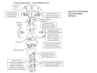
EBV delineating the corticospinal tract. Serviço de Neurorradiologia Hospital Garcia de Orta, E.P.E Lígia Neves ; Ricardo Pimentel, Rui de Carvalho; Miguel Grunho; Cristina Marques; Júlia Duarte. Introduction / Case Report / Discussion / Conclusion.

E N D
EBV delineating the corticospinal tract Serviço de Neurorradiologia Hospital Garcia de Orta, E.P.E Lígia Neves; Ricardo Pimentel, Rui de Carvalho; Miguel Grunho; Cristina Marques; Júlia Duarte
Introduction / Case Report / Discussion / Conclusion • Epstein-Barr virus (EBV) is the pathogen of infeccious mononucleosis, which usually is a benign and self-limited disease. • Although unusual, it may produce neurologic symptoms such as seizures, polyradiculomyelitis, transverse myelitis, encephalitis or cranial nerve palsies.
Introduction / Case Report / Discussion / Conclusion • Male, 40 years old. • Clinical presentation: acute tetraparesis, no reflexes, bilateral D4 sensitive level plus Kernig and Brudzinski signs, dysphagia and no pharyngeal reflex. • No inflammatory blood parameters.
Introduction / Case Report / Discussion / Conclusion 1.5T MRI - bilateral hiperintensity delineating the corticospinal tract Coronal T2 FLAIR Axial T2 FSE
Introduction / Case Report / Discussion / Conclusion 1.5T MRI - hiperintensity delineating the pyramidal tract in the ventral cordbetween segments D2-D10 Axial T2 FSE
Introduction / Case Report / Discussion / Conclusion 1.5T MRI - hiperintensity delineating the corticospinal tract in the ventral spinal cord between D2 and D10. No spinal cord swelling and no parenchymal or meningeal contrast enhancement Sagittal T1 C+ Sagittal T2 FSE
Introduction / Case Report / Discussion / Conclusion • Lombar puncture revealed: • - pleocytosis • - increased protein levels • - decreased glucose levels • Polymerase chain reaction (PCR): • - genomic nucleic acids positive for EBV
Introduction / Case Report / Discussion / Conclusion 1.5T MRI - Partial T2 hiperintensity resolution after treatment with corticotherapy, ceftriaxone, acyclovir and plasmapheresis Coronal T2 FLAIR Sagittal T2 FSE
Introduction / Case Report / Discussion / Conclusion • In this case, the diagnosis of EBV infection was ascertained by PCR. • EBV can be presented with a wide spectrum of imaging abnormalities ranging from normal to diffuse signal intensity changes either in grey or white matter, sometimes resembling ADEM. • The particularity of this case is the distribution of signal intensities confined to the pyramidal tracts.
Introduction / Case Report / Discussion / Conclusion • This case demonstrates that EBV can be presented with a peculiar distribution of MR signal in CNS and therefore should be considered in otherwise unexplained pyramidal tract lesions cases.
References 1.G. Hagemann H.-J. Mentzel H. Weisser A. Kunze C. Terborg , Multiple Reversible MR Signal Changes Caused by Epstein-Barr Virus Encephalitis, AJNR Am J Neuroradiol 27:1447–49, Aug 2006 2. Mascalchi M, Tassinari C. Corticospinal tract degeneration in motor neuron disease, AJNR. 2995; 16: 878-880 3.Volpi A. Epstein-Barr virus and human herpesvirus type 8 infections of the central nervous system. Herpes 2004;11 (suppl 2):120A–27A 4.Fujimoto H, Asaoka K, Imaizumi T, et al. Epstein-Barr virus infections of the central nervous system. Intern Med 2003;42:33– 40