Download
1 / 31
430 likes | 625 Views
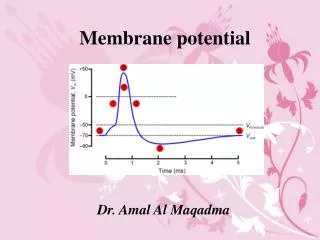
Mitochondrial Membrane Potential (MMP). Contents. Definition of MMP Formation of MMP Measurement/estimation of MMP Role of MMP in function of both normal and pathophysiology. Definition of MMP Formation of MMP

E N D
Contents • Definition of MMP • Formation of MMP • Measurement/estimation of MMP Role of MMP in function of both normal and pathophysiology
Definition of MMP • Formation of MMP • Measurement/estimation of MMP Role of MMP in mitochondrial function of both normal and pathophysiology
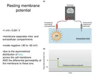
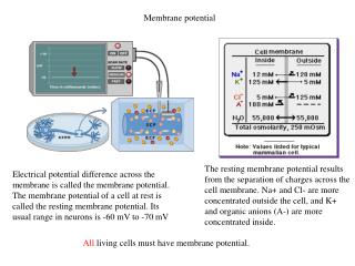
The miotchondrial membrane potential is a highly negative (~ -180mV) potential across the inner membrane of mitochondria.
Definition of MMP • Formation of MMP • Measurement/estimation of MMP Role of MMP in mitochondrial function of both normal and pathophysiology
Sketch map showing the structure of mitochondria (on the left ) and principle of generation of MMP in mitochondria. Electron transport system (ETS) pumps protons across the inner mitochondrial membrane (i.m) and thus generates membrane potential (MMP)
Definition of MMP • Formation of MMP • Measurement/estimation of MMP Role of MMP in mitochondrial function of both normal and pathophysiology
Principle of measurementof MMP • Directly measurement using electrode • Indirectly measurement using fluorescent indicators
Directly measurement using electrode • Indirectly measurement using fluorescent indicators
Directly measurement using electrode • Indirectly measurement using fluorescent indicators
Fluorescent indicators in measurement of MMP • Rhodamine 123 • Tetramethylrhodamine methyl ester (TMRM) • Tetramethylrhodamine ethyl ester (TMRE) • Carbocyanine JC-1
Characteristics of these indicators • Accumulate in the mitochondrial matrix because of the solubility in both the inner mitochondrial membrane and matrix space. • The distribution of these indicators across the inner membrane has been shown to follow the Nernst equation. • Rhodamines are single-excitation, single emission dyes, whereas JC-1 emits red light (590nm) at high concentration (JC-aggregates form), and emits green light (530nm) at low concentrations (monomer form)
Principle of MMP measurement/estimation in isolated ventricular myocytes (1)MMP can be calculated using the following equation : Vm =-(RT/zF) * ln(Fmitochondria/Fnucleus) where Vm is membrane potential, R is the gas constant, T is absolute temperature,F is Faraday’s constant, and F is fluorescence. (2) MMP can be estimated by fluorescence changes in mitochondrial matrix.
Confocal imaging of mitochondria in a guinea pig ventricular myocyte with use of membrane potential indicator tetramethylrhodamine methyl ester (TMRE). Cells were loaded with 200 nM TMRE Bright areas in image represent mitochondria that have preferentially accumulated TMRE. Brightest mitochondria are those centered within the focal plane. Fluorescence detection • (1)Confocal fluorescence microscopy
Laser scanning confocal microscopic images of JC-1(1ug/ml) fluorescenceA: Control astrocytes. B: NH4Cl treatment showing a significant degree of depolarization. C:Cells treated with NH4Cl + CsA(5 mM) are similar to control. FCCP (3 mM, 20 min) induced a total collapse of the Dcm (inset in A). Graph on the left shows the time-dependence of ammonia (5 mM ) induced dissipation of the MMP in atrocytes. Quantitation is expressed as mean red/green fluorescence ratios and compared to untreated controls that were set at 100%. *p<0.05 vs control.
(2) Flow cytometre Effects of 48hr with 5 mM NH4Cl on the MMP as determined by flow cytometry.Data is plotted as cell counts vs. TMRE fluorescence.ammonia results in a spectral shift to the left indicating mitochondrial depolarization. This effect was blocked by CsA (5 uM).A:Control. B: 5 mM NH4Cl .C: 5 uM CsA.D: NH4Cl+ CsA. E: FCCP
Definition of MMP • Formation of MMP • Measurement/estimation of MMP Role of MMP in mitochondrial function of both normal and pathophysiology
IN normal cell function of MMP • Maintenance of MMP is essential for ATP synthesis, i.e. providing motive force for translocatione of proton from mitochondrial matrix into the intermembrane space. • MMP provides driving force for Ca2+ uptake into mitochondria by the Ca2+ uniporter.
Maintenance of MMP is essential for ATP synthesis, i.e. providing motive force for translocatione of proton from mitochondrial matrix into the intermembrane space. • MMP provides driving force for Ca2+ uptake into mitochondria by the Ca2+ uniporter.
The MMP is necessary for conversion of ADP to ATP (ATP synthesis).
Maintenance of MMP is essential for ATP synthesis, i.e. providing motive force for translocatione of proton from mitochondrial matrix into the intermembrane space. • MMP provides driving force for Ca2+ uptake into mitochondria by the Ca2+ uniporter.
PTP = permeability transition pore, mNCE = mitochondrial sodium calcium exchanger, F1F0 = ATP synthase, e.t.c = electron transport chain, mCU = mitochondrial calcium uniporter, DpH is the pH gradient (~0.5) and DY is the membrane potential (~180mV)
MMP in pathophysiology • MMP in oxygen stress • MMP in ischemia/reperfusion • MMP in IPC
MMP in oxygen stress • MMP in ischemia/reperfusion injury • MMP in IPC
Mitochondrial defence against radical-induced oxidative stress. A: under normal conditions, respiratory chain (RC) produces 2~4% reactive oxygen species (ROS)from an electron flux. B: higher cellular oxygen causes increase of ROS, and radical scavengers is the first step to defense.C:if scavengers are exausted, the increased oxygen stress result in an increase of proton conductance and a small dissipation of MMP.D: PTP opening when a certain MMP decline is reached. E:ongoing oxidative stress resulted in irreversible permeability transition causing a breakdown of MMP, cytochrome c release,cell apoptosis.
MMP in oxygen stress • MMP in • MMP in ischemia/reperfusion injury • MMP in IPC
Time course of changes in cell length and mitochondrial membrane potential during anoxia and reoxygenation.
MMP in oxygen stress • MMP in ischemia/reperfusion injury • MMP in IPC
It has been reported that the mitochondrial ATP- sensitive potassium channel play important role in the cardioprotection of IPC Nicorandil, a selective mKATP opener partially depolarized the MMP