Download
1 / 29
320 likes | 424 Views
American Trypanosomiasis (Chagas disease). Trypanosoma cruzi belongs to the subkingdom Protozoa. They are flagellar organisms that have one nucleus and an organelle, the kinetoplast, that gives rise to one mitochondrion and mitochondrial DNA. Classification.

E N D
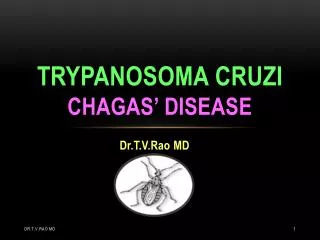
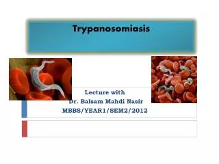
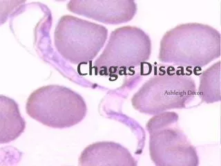
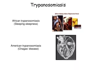
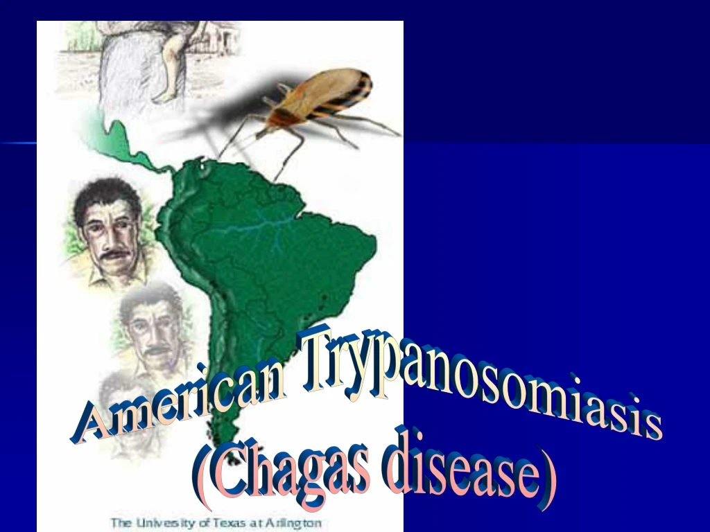
American Trypanosomiasis (Chagas disease)
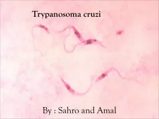
Trypanosoma cruzi belongs to the subkingdom Protozoa. They are flagellar organisms that have one nucleus and an organelle, the kinetoplast, that gives rise to one mitochondrion and mitochondrial DNA.
Classification • Eukaryota (organisms with nucleated cells),Kingdom Protista, Phylum Protozoa. • Trypanosoma cruzi. • T. cruzi reproduce asexually by binary fission. • Like all other trypanosomes, T. cruzi live one stage of their lives in the blood and/or tissues of vertebrate hosts and during other stages they live in the digestive tracts of invertebrate vectors (temporary hosts).
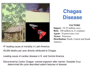
Chagas disease, named for Carlos Chagas, the Brazilian doctor who first described the disease in 1909, is caused by Trypanosoma cruzi,a flagellate protozoan parasite. Case of human trypanosomiasis have been reported in almost all countries of the Americas, including the southern United States, but the main foci are in poor rural areas of Latin America.
Life cycle of Trypanosoma cruzi. • An infected triatomine insect vector (or “kissing” bug) takes a blood meal and releases trypomastigotes in its feces near the site of the bite wound. • Inside the host, the trypomastigotes invade cells, where they differentiate into intracellular amastigotes . • The amastigotes multiply by binary fission and differentiate into trypomastigotes, and then are released into the circulation as bloodstream trypomastigotes
Trypomastigotes infect cells from a variety of tissues and transform into intracellular amastigotes in new infection sites. • The bloodstream trypomastigotes do not replicate. • Replication resumes only when the parasites enter another cell or are ingested by another vector. • The “kissing” bug becomes infected by feeding on human or animal blood that contains circulating parasites. • The ingested trypomastigotes transform into epimastigotes in the vector’s midgut . • The parasites multiply and differentiate in the midgut.
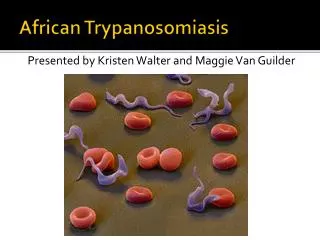
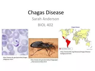
Epidemiology • Chagas disease is transmitted by cone-nosed triatomine bugs of several genera (Triatoma, Rhodnius, Panstrongylus). • Trypanosoma cruzi can also be transmitted through blood transfusions, organ transplantation, transplacentally, breast milk and in laboratory accidents.
Transmission Methods of T. cruzi • Contamination Contamination through the insect's feces is the primary mechanism by which vinchucas pass T. cruzi to humans.
Transmission Methods of T. cruzi • Blood Transfusions and Organ Transplants Blood transfusions are the second most common mechanism of transmission of Chagas' disease to people in Latin America, Europe,and the United States.
Transmission Methods of T. cruzi • Transmission Through Birth Mothers pass T. cruzi on to their children as T. cruzi travels through the placenta, birth canal, and possibly maternal milk. This type of transmission occurs less frequently than other methods. Possibilites include diffusion of the parasite across the extra-embryonic membranes, or through the maternal blood supply.
Geographic • Chagas disease is found only in Latin America
Natural foci of Chagas disease exist among wild mammals and their associated triatomines. • Humans and domestic animals became involved in the epidemiologic chain several centuries ago, when insects living under wild conditions began adapting to households. • Opossums, armadillos, and wild rodents are reservoirs of the parasite, linking the wild and domestic cycles
Pathogenesis Inflammatory reactions at the sites of rupturing pseudocysts can lead to pathologic manifestations, such as acute myocarditis and destruction of parasympathetic ganglia of the heart and myenteric plexus. An autoimmune reaction may develop.
Clinical Symptoms. The incubation period is 7-14 days. The humandisease occurs in 3 stages: • the acute stage shortly after the infection; • the indeterminate stage; • the chronic stage that may develop over 10 years.
Acute phaseof Chagas disease • A local skin nodule called a chagoma can appear at the site of inoculation. • When the inoculation site is the conjunctival mucous membranes, the patient may develop unilateral periorbital edema, conjunctivitis, and preauricular lymphadenitis. (Romaña's sign). • The acute phase is usually asymptomatic, but may present symptoms of fever, anorexia, lymphadenopathy, mild hepatosplenomegaly, and myocarditis.
Other symptoms are: • tiredness, • sometimes a rash, • loss of appetite, diarrhea, and vomiting. • Infants and very young children can get an often-fatal swelling of the brain.
Indeterminate stage • During the indeterminate stage, about 8 to 10 weeks after infection, infected persons have no symptoms.
Chronic stage of Chagas disease The disease affects the nervous system, digestive system and heart: • dementia, • damage to the heart muscle (cardiomyopathy), altered heart rate or rhythm, • sometimes dilation of the digestive tract (megacolon and megaesophagus), • Weight loss. • Swallowing difficulties may be the first symptom of digestive disturbances and may lead to malnutrition. • Left untreated, Chagas disease can be fatal, in most cases due to the cardiomyopathy component.
The risk factors for gettingChagas disease • International travel • Undeveloped countries • Poorly constructed houses • Rural areas • Mud houses • Adobe houses • Thatch houses • Assassin bugs • Persons with weakened immune systems are at risk of severe infections and complications.
Laboratory Diagnostics • microscopic blood examination, • Xenodiagnosis; • by culturing the blood. • serologic tests : • indirect hemagglutination, • indirect immunofluorescence, • enzyme-linked immunosorbent assay (ELISA)]
Xenodiagnosis • In this test, uninfected vinchucas are placed in a jar and tucked under the armpit of a patient suspected of being infected. • The vinchucas are allowed to consume blood for thirty minutes, and their feces are examined for T. cruzi thirty and sixty days afterward. • This technique is rarely used on children, and many adults have are hesitant in being willfully bitten by vinchucas.
ELISA testing • People without noticeable signs of chagas who live in chagasic areas should be encouraged to have an ELISA test.
Culturing the blood. Trypanosoma cruzi in cultured HeLa cells (Giemsa) Amastigotes infecting cells of muscle tissue
Treatment • No effective treatment. • Available drugs only kill extracellular parasites. • Benznidazole and Nifurtinox: current drugs of choice. Required daily for up to 2 months or more. • Hospitalization may be needed because of adverse effects
Preventation • There is no vaccine or drug to prevent Chagas disease. When traveling to areas where Chagas disease occurs, follow these precautions: • Avoid sleeping in poorly constructed thatch, mud, or adobe houses. If that is not possible, use a bednet. • Use insecticides to kill insects and reduce the risk of transmission. • Be aware of the risk of contracting Chagas disease through blood transfusions. In many countries, the blood supply is not well screened.