Download
1 / 37
380 likes | 456 Views
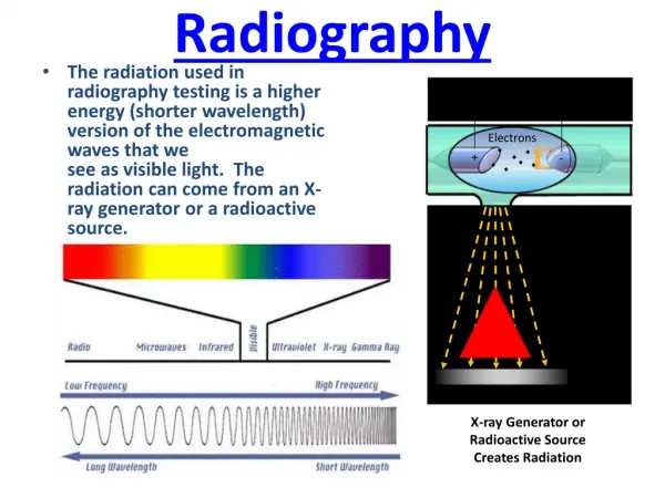
Radiography. What are some ways we can see inside of something?. X-Ray Imaging. Electromagnetic radiation between Ultraviolet and Gamma rays Produced by accelerated electrons colliding with tungsten anode and projected at body part

E N D
X-Ray Imaging • Electromagnetic radiation between Ultraviolet and Gamma rays • Produced by accelerated electrons colliding with tungsten anode and projected at body part • Calcium in bones and added contrasts of barium or iodine absorb x-rays • Image receptor digital films reads black where x-rays penetrated through body
MRI • Machine creates a magnetic field that aligns the magnetization of some atoms in the body and radio frequency fields further alter the alignment. Nuclei produce rotation magnetic field, which is detected by the scanner • Primarily used to examine soft tissue in body, brain, muscles, heart and cancers • No ionizing radiation
Ultrasound • Cyclic sound pressure with frequency greater than 20,000 hertz • Transducer sends and receives sound signals in medium • Used by bats for hunting • Medical sonography used for soft tissue diagnosis, fetuses, interventional procedures
CT Scan • X-ray Computed Tomography (CT) • Tomography-means imaging by sections from penetrating wave • X-ray source rotates helically about patient or specimen to create 3D image from sectioning • Useful for preventative medicine
PET Scan • Positron Emission Tomography • Positron - antielectron (positive electron) • System uses a tracer (positron-emitting radionuclide) to detect gamma rays emitted by the collision of these positrons with electrons • Fludeoxyglucose (sugar) is commonly used as tracer
Attenuate Means to reduce or weaken The higher the attenuation coefficient the greater the ability of the material to reduce or weaken the radiation beam This can be done by scattering, absorption What attenuates x-rays during medical procedures?
Compare Different Materials Cadmium ( Cd ) Lead ( Pb ) Plastic ( (CH3)n)
Which will block the radiation? Yes Yes Yes No No Yes
Neutron Imaging System Video output Gamma rays (photons) electrons neutrons Reactor D2O Tank Phosphorescent screen (similar to the one in your TV) converts electrons to visible light. Scintillation material (GdO2S)converts neutrons into gamma rays through neutron capture reactions Photocathode coverts photons to electrons through photoelectric effect Neutron Imaging System “3.5 hours slide” courtesy of Danielle Hauck.5/17/2004- rev. 9/29/04 CCD
CODE Box Student Project to Demonstrate X-Ray/Neutron Radiography Was originally in cardboard shoe box, but was replaced by more durable aluminum.
Cadmium C O D E Lead
Cadmium C O D E Lead Plastic P S U
Hydrogen Fuel Cell Imaging Fuel Cell research conducted at RSEC
Hydrogen Fuel Cell Imaging Water Calibration Wedge
Figure it out • Look at the next slide and see if you can figure out what will show up on x-rays
Cad Lead Plastic
Figure it out neutrons • Look at the picture again and figure out what you will see with neutrons.
Cad Lead Plastic
Cadmium Lead Plastic
Cad Lead Plastic
WSHI design project • Design a project using cadmium, lead and plastic that will look different with x-rays and neutrons • Table shows what material blocks the radiation
Resources • Images courtesy of WikiMedia Commons and Breazeale Nuclear Reactor • Hubbell, J. H. "NIST: X-Ray Mass Attenuation Coefficients." National Institute of Standards and Technology. Web. 17 Sept. 2011. <http://www.nist.gov/pml/data/xraycoef/index.cfm>. • Whaites, Eric; Roderick Cawson (2002). Essentials of Dental Radiography and Radiology. Elsevier Health Sciences. pp.ハ15ミ20. ISBNハ044307027X. • Special Thanks to: Joanna Kinney and Jeremiah Lynch for preparing this presentation at Penn State University, Radiation Science & Engineering Center