Download
1 / 47
661 likes | 1.79k Views
Polydiacetylene Vesicles: Direct Biosensors with a Colorimetric Response. Margaret A. Schmitt Samuel H. Gellman Group University of Wisconsin, Madison February 20, 2003. Outline. Biosensors Definition and Introduction Direct and Indirect Polydiacetylenes Polymerization Reaction

E N D
Polydiacetylene Vesicles: Direct Biosensors with a Colorimetric Response Margaret A. Schmitt Samuel H. Gellman Group University of Wisconsin, Madison February 20, 2003
Outline • Biosensors • Definition and Introduction • Direct and Indirect • Polydiacetylenes • Polymerization Reaction • Chromic Response to Environmental Changes • Harnessing Chromic Response in a Useful Biosensor Construct • Variables Associated with Designing an Appropriate PDA Biosensor for a Variety of Systems
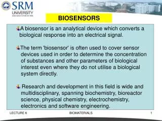
Biosensors • Device incorporating a biological sensing element directly connected to a signal transducer • Biosensor design research attempts to couple Nature’s “lock-and-key” interactions with cleverly engineered signal transduction mechanisms • Molecular recognition assumes many forms: enzyme-substrate, antibody-antigen, and receptor-ligand interactions • Two-fold utility: • Basic science level: develops an understanding of complex biological processes • Applied science level: broad applicability in industrial and medicinal settings Analyte “recognition” Signal Biologically Sensitive Element X No interaction No signal
Importance of Biosensing Techniques • Glucose level monitoring in individuals with diabetes • Rapid detection of toxins and other biological warfare agents 1 2 4 3 http://www.healthchecksystems.com/bioscanner.htm
Designing Usable Biosensors • Signal must have a direct relationship to quantity of material being analyzed • Sensor must demonstrate specificity and selectivity in recognizing a single compound or group of compounds in a varied mixture
Biosensor Detection Ranges • Detection limit of sensor must be within a relevant range • Sensor must have a reasonable response time
Indirect vs. Direct Biosensors Indirect: Relies on detection of a labeled ligand after a binding event has occurred. Direct: Binding event is directly linked to a signal transduction event for detection in real-time.
Indirect Biosensor: ELISA Wash Incubate Wash Incubate • Based upon tight binding between an antigen and antibody • Labeling agents for ligands include fluorescent probes and radioisotopes • Most commonly used reporter enzyme horseradish peroxidase (HRP), which upon reaction with substrate produces a bright green color
Direct Biosensor: SPR • Optical detection method for studying interactions between a soluble analyte and immobilized ligand • Binding of the analyte molecule changes the refractive index in a way that is approximately proportional to the mass of the molecules which have entered the interface • Stoichiometry of binding can be examined http://www.astbury.leeds.ac.uk/Facil/spr.htm
Indirect Advantages Amount of material required Can detect virtually any material (ELISA) Sensitivity Signal amplification Disadvantages Labeled ligands or secondary reagents required Background problems – washing is necessary Direct Advantages Binding event results in signal transduction Signal measures only the desired interaction Disadvantages Specialized machinery is often required Signals more difficult to amplify Time-consuming Advantages and Disadvantages of Indirect and Direct Biosensors
“Ideal” Biosensors • Response is directly coupled to recognition event: Direct • Signal readily detectable without the use of expensive or large instrumentation • Adaptable to detect many types of analytes
Outline • Biosensors • Definition and Introduction • Direct and Indirect • Polydiacetylenes • Polymerization Reaction • Chromic Response to Environmental Changes • Harnessing Chromic Response in a Useful Biosensor Construct • Variables Associated with Designing an Appropriate PDA Biosensor for a Variety of Systems
Diacetylene Polymerization • Topochemical polymerization reaction • Reaction is very sensitive to the surrounding environment and packing of substituents • Reacting carbon atoms must be less than 4 Å away from each other or polymerization is not likely to occur
No butatrienic structure indicated in either blue or red form as indicated by 13C NMR Carbon Blue Phase (ppm) Red Phase (ppm) >C= 131.6 132.0 −C≡ 107.4 103.6 Tanaka, H.; et. al.Macromolecules1989, 22, 1208. PDA Response to Environmental Changes
Carbon Blue (ppm) Red (ppm) δ-CH2 66.6 65.5 α-CH2 37.3 37.8 ε-CH2 32.9 32.6 β,γ-CH2 24.5 26.4 Effect of Side Chain Conformation on Chromatic Response • Only β,γ-carbons show significant shift between 2 phases • Conformational change around backbone single bonds is minimal as α-carbon chemical shifts to not change significantly Tanaka, H.; et. al.Macromolecules1989, 22, 1208.
Polydiacetylenes as Biosensors • Incorporation into vesicles • Methodology of assay • Physical changes in vesicles and relationship to color change • Variables associated with appropriate biosensor design • Position of diacetylenic functionality • Incorporation of recognition element ?
Two Supramolecular Approaches for Utilizing Polydiacetylenes in Sensors • Immobilization of polymer as a • thin film on a solid glass support Solution-based sensors incorporating PDA vesicles (liposomes)
Advantages of Vesicles • Liposomes can be made more simply and reproducibly • Vesicle assays and analysis can be done in a 96-well plate format • Liposomes mimic the cell membrane more closely than thin films • Ability to immobilize and remain functional on a surface
Vesicle Immobilization onto Au Films • Use lipid with disulfide containing headgroup to immobilize vesicles on gold • Disulfide remains oxidized, reducing vesicle aggregation • Vesicles remain highly monodisperse over periods of 3 days in buffer Stanush, I.; Santos, J.; Singh A. J. Am. Chem. Soc.2001, 123, 1008.
PDA Vesicle Stability • Stable at 4ºC in solution for months • Can be made stable to lyophilization and resuspension • Not sensitive to white light • Show no evidence of fusion to form large aggregates • Not destroyed osmotically by salts
PDA Vesicles: Synthesis • Diacetylenic monomer molecules self-assemble into an ordered array by the same driving forces which occur in the formation of biological membranes • Vesicle formation is encouraged by sonication • Monitor polymerization reaction by appearance of a deep blue color
Design of Colorimetric Assay • Analyze peptide-membrane interactions • Utilize well-characterized antimicrobial peptides and related mutants to examine interactions at vesicle surface and colorimetric response (CR) • Amphiphilic peptides severely disrupt membrane surface and may insert into membrane and form pores • Vesicles contained 6:4 mole ratio of TRCDA and phospholipid (e.g. DMPC) Kolusheva, S.; Boyer, L.; Jelinek, R. Nature Biotechnology2000, 18, 225. Kolusheva, S.; Shahal, T.; Jelinek, R. Biochemistry2000, 39, 15851.
Colorimetric Assay • Vesicle solutions buffered with Tris to pH 8.5 • Incubate peptide and vesicles for 30 min at 27ºC and measure CR Control: no peptide Cells containing amphiphilic peptides Kolusheva, S.; Boyer, L.; Jelinek, R. Nature Biotechnology2000, 18, 225. Kolusheva, S.; Shahal, T.; Jelinek, R. Biochemistry2000, 39, 15851.
Calculation of quantitative value for extent of color transition from initial blue state to final red state A: absorbance at the “blue” (~640nm) or “red” (~500nm) Depending upon background levels and non-specific interactions, interactions can be detected with as little as 5-7% CR Colorimetric Response (CR) f 0 0 Kolusheva, S.; Boyer, L.; Jelinek, R. Nature Biotechnology2000, 18, 225. Kolusheva, S.; Shahal, T.; Jelinek, R. Biochemistry2000, 39, 15851.
Non-Specific PDA-Analyte Interactions • Measure CR with pure PDA vesicles to determine changes due to interactions between analyte and negatively charged PDA portion of vesicles • Even at μM concentrations, melittin can be detected above background Kolusheva, S.; Boyer, L.; Jelinek, R. Nature Biotechnology2000, 18, 225. Kolusheva, S.; Shahal, T.; Jelinek, R. Biochemistry2000, 39, 15851.
Negative Controls • Expose vesicles to mismatched analyte to rule out CR resulting from non-specific interactions with the recognition element • Examine response due to presence of peptides not expected to be membrane active (e.g. neuropeptides) • Peptide-membrane interactions are non-specific; use to ensure CR is due only to membrane interactions and disruption and not presence of other analytes No peptide Antimicrobial Peptides Neuropeptide (no membrane interaction) Kolusheva, S.; Boyer, L.; Jelinek, R. Nature Biotechnology2000, 18, 225. Kolusheva, S.; Shahal, T.; Jelinek, R. Biochemistry2000, 39, 15851.
Polydiacetylenes as Biosensors • Incorporation into vesicles • Methodology of assay • Physical changes in vesicles and relationship to color change • Variables associated with appropriate biosensor design • Position of diacetylenic functionality • Incorporation of recognition element ?
Effective length of conjugation in the polymer shortens as a result of desired interaction resulting in a strong blue-red color transition Mechanisms of Biochromatic Response • Insertion of viral membrane or toxin hydrophobic domains into the PDA bilayer • Multipoint interactions of the receptor at the PDA-vesicle surface changing packing of lipid headgroups near surface
Observation of Physical Changes in Vesicles in Conjunction with CR • Detection of antibody-epitope recognition • HA peptide-epitope is presented at the N-terminus of a hydrophobic -helix designed to span lipid bilayers Kolusheva, S.; et. al. J. Am. Chem. Soc.2001, 123, 417.
Prior to addition of antibody Incubated with HA antibody Incubated with incorrect antibody Antibody-Epitope Interaction Results in a Physical Change Vesicles Blue Vesicles Red Vesicles Blue Kolusheva, S.; et. al. J. Am. Chem. Soc.2001, 123, 417.
PLA2 PLC PLD Varied Mechanisms of Membrane Interaction • Evidence of phospholipase activity : PLA2, PLC, and PLD • Enzymes which hydrolyze cell membrane phospholipids • Each enzyme cleaves PC in a different location, but activity of each results in a similar colorimetric response Jelinek, R.; et. al. Chem. Biol.1998, 5, 619. Okada, S.; Jelinek, R.; Charych, D. Angew. Chem. Int. Ed.1999, 38, 655.
Cleavage Products Disrupt Membrane • PLA2 (acyl hydrolase) • Cleavage products leave membrane matrix • Forms “pits” in membrane surface, resulting in changes in lipid packing • PLC (phosphodiesterase) • Cleavage product is 1,2-diacylglycerol • Lipid chains spread apart and expose hydrocarbon core to aqueous surface • PLD (phosphodiesterase) • Cleavage product is phosphatidic acid (PA) • PA has affinity for Ca2+ ions in buffer • Interaction with cations results in vesicle condensation Jelinek, R.; et. al. Chem. Biol.1998, 5, 619. Okada, S.; Jelinek, R.; Charych, D. Angew. Chem. Int. Ed.1999, 38, 655.
Polydiacetylenes as Biosensors • Incorporation into vesicles • Methodology of assay • Physical changes in vesicles and relationship to color change • Variables associated with appropriate biosensor design • Position of diacetylenic functionality • Incorporation of recognition element ?
Location of Polymerization Group • 10,12-PDAs have a much more rigid hydrophobic chain prior to the diacetylene moiety • Strong connection between conformation of alkyl chain and polymer electronic properties • 5,7-PDAs are expected to be more responsive to environmental changes m = 0 or 6 m = 0: TCDA and DCDA m = 6: TRCDA and PCDA
Thermochromism of 5,7- and 10,12-PDAs • Examine thermochromism in response to incubation at 50ºC as a function of time • Vesicles composed of 5,7-PDAs express an enhanced response compared to 10,12-PDAs • Drawback of this enhanced response is that 5,7-PDAs are more readily affected by properties of their solution: salt content, pH, etc Okada, S.; et. al. Acc. Chem. Res. 1998, 31, 229.
Positive response to E. coli with 2,4-PDA vesicles (and sialic acid receptor) No response to E. coli with 10,12-PDA vesicles Positive response to cholera toxin with 5,7-PDA vesicles (and ganglioside receptor) No response to cholera toxin with 10,12-PDA vesicle Location of Polymer Backbone and Effective Biochromic Response vs. vs. Pan, J.; Charych, D. Langmuir 1997,13, 1365. Ma, Z.; et. al.J. Am. Chem. Soc. 1998, 120, 12678.
Incorporated on separate membrane-spanning peptide in antibody-epitope studies Synthetically attach recognition element to lipid containing diacetylene moiety Incorporate recognition element through a lipid in the system which does not contain a diacetylene moiety, and therefore cannot be polymerized Incorporation of Recognition Element
Synthetic Attachment of Recognition Element • Bifunctional molecule incorporates both the recognition element (sialic acid) and the reporter diacetylene moiety • Surface lectin of influenza virus (hemagglutinin) binds terminal -glycosides (sialic acid residues) on cell surface glycoproteins and glycolipids 10,12-pentacosadiynoic acid (PCDA) Reichert, A.; et. al. J. Am. Chem. Soc. 1995,117, 829.
PDA Vesicle Detection of Influenza Virus • HA binds cell surface sialic acid residues and initiates viral infection • Detection of as little as 11 HAUs of virus particle (~11 x 107 virus particles) Reichert, A.; et. al. J. Am. Chem. Soc. 1995,117, 829.
Incorporation of Recognition Element on a Non-Polymerizable Lipid • Useful when receptor of interest is already lipid linked or when attaching receptor to diacetylenic lipid may be synthetically challenging • Gangliosides are lipid molecules that reside on the surface of the cell membrane and display carbohydrate recognition groups • Cholera toxin recognizes GM1 ganglioside 5,7-docosadiynoic acid (DCDA) Pan, J.; Charych, D. Langmuir 1997,13, 1365.
Detection of Cholera Toxin • Detection of slightly less than 100 μg/ml cholera toxin • Response is slightly sigmoidal • Binding cooperativity – binding one ligand makes the vesicle more accessible for others • Polymer side chain conformations – once the effective conjugated length of the vesicle is perturbed as the result of toxin binding, subsequent perterbation is more favorable Pan, J.; Charych, D. Langmuir 1997,13, 1365.
Screening a Library with PDA Vesicles • Examine structure-activity relationships in a library of amphiphilic co-polypeptides • Relationship between polypeptide -amino acid composition and interaction with phospholipids found in cell membranes • Suggest important factors for designing new antimicrobial peptides Wyrsta, M.; Cogen, A.; Deming, T. J. Am. Chem. Soc.2001, 123, 12919.
Ala, Phe, and Leu: α-helix favoring Ile and Val: β-sheet favoring Detection of Peptide-Membrane Interactions • Most membrane-active peptides are of intermediate chain length and high hydrophobic content • Peptides containing -helix favoring amino acids interact with vesicles and produce a colorimetric response • Peptides containing β-sheet favoring amino acids do not produce any colorimetric response B) Lys/Ala peptides E) Lys/Ile peptides C) Lys/Phe peptides F) Lys/Val peptides D) Lys/Leu peptides Blue = negative Red/Orange = positive Wyrsta, M.; Cogen, A.; Deming, T. J. Am. Chem. Soc.2001, 123, 12919.
Future Directions • Continue to examine the mechanism of PDA biochromic response • Apply vesicle methodology in evaluation of compounds with unknown activity (e.g. potential antimicrobial peptides or enzyme inhibitors) • Correlate colorimetric response with desired biological interaction • Examine biochromic responses in new constructs and immobilized vesicles ?
Conclusions • Polydiacetylene vesicles mimic the properties of cell signaling by directly coupling a bio-recognition event to signal transduction • Recognition events in a PDA vesicle result in a visible colorimetric signal which changes from blue to red • PDAs are able to detect peptide-membrane interactions, antibody-epitope recognition, enzyme binding and catalysis, and virus and toxin molecule recognition • If a relationship between the colorimetric response of PDAs and desired bio-recognition events can be shown, PDA vesicles could become a useful sensing technique with a wide variety of applications
Acknowledgements Gellman Group Nick Fisk Terra Potocky Tim Peelen Jon Lai Marissa Rosen Erin Carlson