Download
1 / 17
170 likes | 254 Views
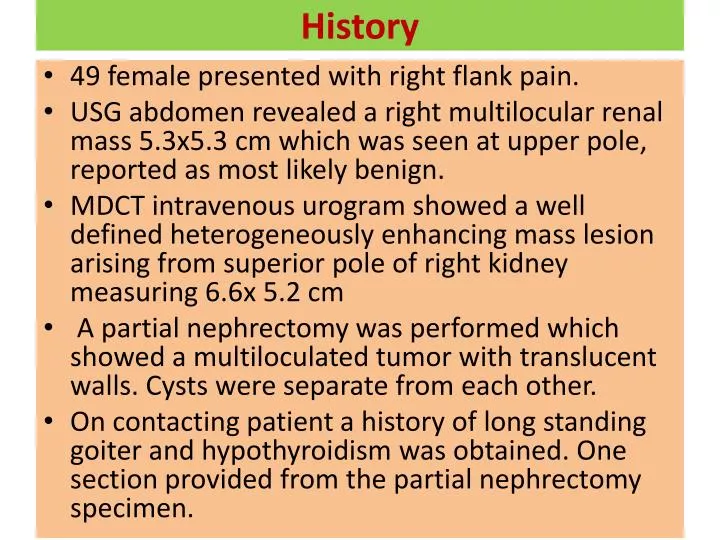
History. 49 female presented with right flank pain. USG abdomen revealed a right multilocular renal mass 5.3x5.3 cm which was seen at upper pole, reported as most likely benign.

E N D
History • 49 female presented with right flank pain. • USG abdomen revealed a right multilocular renal mass 5.3x5.3 cm which was seen at upper pole, reported as most likely benign. • MDCT intravenous urogram showed a well defined heterogeneously enhancing mass lesion arising from superior pole of right kidney measuring 6.6x 5.2 cm • A partial nephrectomy was performed which showed a multiloculated tumor with translucent walls. Cysts were separate from each other. • On contacting patient a history of long standing goiter and hypothyroidism was obtained. One section provided from the partial nephrectomy specimen.
? Diagnosis ?? Differential diagnosis
AMACR- internal control AMACR-negative EMA -negative EMA- internal control