Download
1 / 1
10 likes | 114 Views
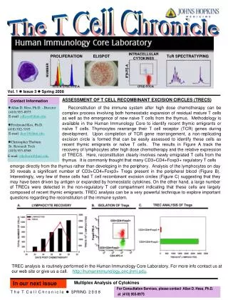
The T Cell Chronicle. Vol. 1 Issue 4 Spring 2009. ANTIGEN-SPECIFIC T-CELL TRACKING UTILIZING THE V b -CDR3 DOMAIN

E N D
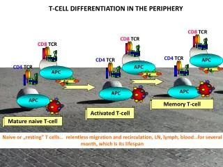
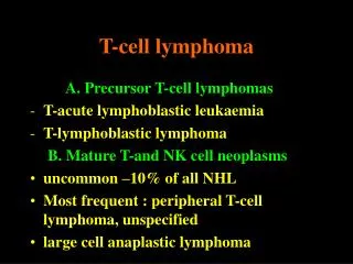
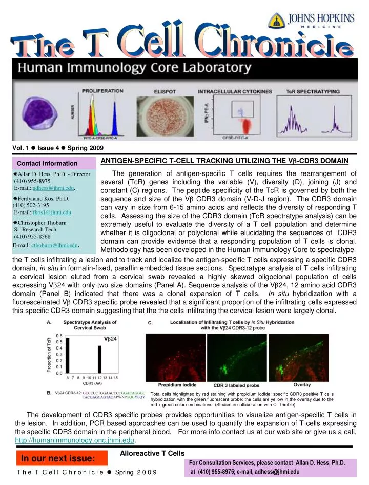
The T Cell Chronicle Vol. 1 Issue 4 Spring 2009 ANTIGEN-SPECIFIC T-CELL TRACKING UTILIZING THE Vb-CDR3 DOMAIN The generation of antigen-specific T cells requires the rearrangement of several (TcR) genes including the variable (V), diversity (D), joining (J) and constant (C) regions. The peptide specificity of the TcR is governed by both the sequence and size of the Vb CDR3 domain (V-D-J region). The CDR3 domain can vary in size from 6-15 amino acids and reflects the diversity of responding T cells. Assessing the size of the CDR3 domain (TcR spectratype analysis) can be extremely useful to evaluate the diversity of a T cell population and determine whether it is oligoclonal or polyclonal while elucidating the sequences of CDR3 domain can provide evidence that a responding population of T cells is clonal. Methodology has been developed in the Human Immunology Core to spectratype Contact Information Allan D. Hess, Ph.D. - Director (410) 955-8975 E-mail: adhess@jhmi.edu. Ferdynand Kos, Ph.D. (410) 502-3195 E-mail: fkos1@jhmi.edu. Christopher Thoburn Sr. Research Tech (410) 955-8568 E-mail:cthoburn@jhmi.edu. - the T cells infiltrating a lesion and to track and localize the antigen-specific T cells expressing a specific CDR3 domain, in situ in formalin-fixed, paraffin embedded tissue sections. Spectratype analysis of T cells infiltrating a cervical lesion eluted from a cervical swab revealed a highly skewed oligoclonal population of cells expressing Vb24 with only two size domains (Panel A). Sequence analysis of the Vb24, 12 amino acid CDR3 domain (Panel B) indicated that there was a clonal expansion of T cells. In situ hybridization with a fluoresceinated Vb CDR3 specific probe revealed that a significant proportion of the infiltrating cells expressed this specific CDR3 domain suggesting that the the cells infiltrating the cervical lesion were largely clonal. Total cells highlighted by red staining with propidium iodide; specific CDR3 positive T cells hybridization with the green fluorescent probe; the cells are yellow in the overlay due to the red + green color combinations. (Studies in collaboration with C. Trimble) The development of CDR3 specific probes provides opportunities to visualize antigen-specific T cells in the lesion. In addition, PCR based approaches can be used to quantify the expansion of T cells expressing the specific CDR3 domain in the peripheral blood. For more info contact us at our web site or give us a call. http://humanimmunology.onc.jhmi.edu. Alloreactive T Cells In our next issue: For Consultation Services, please contact Allan D. Hess, Ph.D. at (410) 955-8975; e-mail, adhess@jhmi.edu T h e T C e l l C h r o n i c l e Spring 2 0 0 9