Download
1 / 69
690 likes | 769 Views
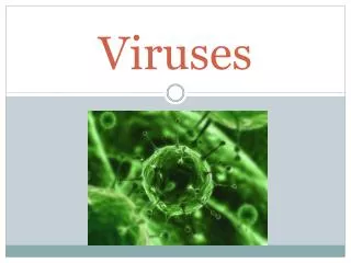
Viruses. 0. 17. Overview: A Borrowed Life. A virus is an infectious particle consisting of little more than genes packaged into a protein coat Viruses lead “ a kind of borrowed life, ” existing in a shady area between life-forms and chemicals. Figure 17.1. 0.25 m.

E N D
Viruses 0 17
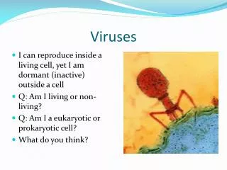
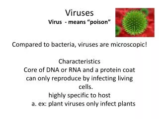
Overview: A Borrowed Life A virus is an infectious particle consisting of little more than genes packaged into a protein coat Viruses lead “a kind of borrowed life,” existing in a shady area between life-forms and chemicals
Figure 17.1 0.25 m
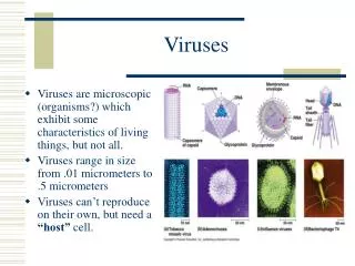
Concept 17.1: A virus consists of a nucleic acid surrounded by a protein coat Even the largest known virus is barely visible under the light microscope Some viruses can be crystalized Viruses are not cells but are a nucleic acid enclosed in a protein coat
Viral Genomes Viral genomes may consist of either Double- or single-stranded DNA, or Double- or single-stranded RNA Depending on its type of nucleic acid, a virus is called a DNA virus or an RNA virus
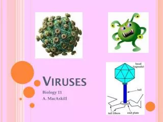
Capsids and Envelopes A capsid is the protein shell that encloses the viral genome Capsids are built from protein subunits called capsomeres A capsid can have various structures
Figure 17.2 Membranous envelope Capsomere RNA RNA DNA Head DNA Capsid Capsomereof capsid Tailsheath Tailfiber Glycoprotein Glycoproteins 80 225 nm 70–90 nm (diameter) 80–200 nm (diameter) 18 250 nm 20 nm 50 nm 50 nm 50 nm (c) Influenza viruses (a) Tobacco mosaic virus (b) Adenoviruses (d) Bacteriophage T4
Figure 17.2a Capsomere RNA DNA Capsomereof capsid Glycoprotein 70–90 nm (diameter) 18 250 nm 20 nm 50 nm (b) Adenoviruses (a) Tobacco mosaic virus
Figure 17.2aa 20 nm (a) Tobacco mosaic virus
Figure 17.2ab 50 nm (b) Adenoviruses
Figure 17.2b Membranous envelope RNA Head DNA Capsid Tailsheath Tailfiber Glycoproteins 80–200 nm (diameter) 80 225 nm 50 nm 50 nm (c) Influenza viruses (d) Bacteriophage T4
Figure 17.2ba 50 nm (c) Influenza viruses
Figure 17.2bb 50 nm (d) Bacteriophage T4
Some viruses have membranous envelopes that help them infect hosts These viral envelopes are derived from the host cell’s membrane and contain a combination of viral and host cell molecules
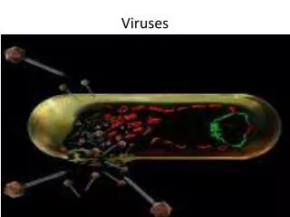
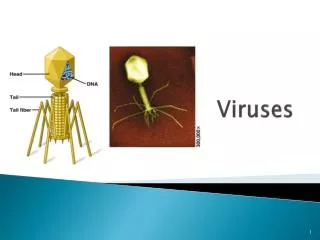
Bacteriophages, also called phages, are viruses that infect bacteria They have the most complex capsids found among viruses Phages have an elongated capsid head that encloses their DNA A protein tail piece attaches the phage to the host and injects the phage DNA inside
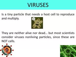
Concept 17.2: Viruses replicate only in host cells Viruses are obligate intracellular parasites, which means they can replicate only within a host cell Each virus has a host range, a limited number of host cells that it can infect
General Features of Viral Replicative Cycles Once a viral genome has entered a cell, the cell begins to manufacture viral proteins The virus makes use of host enzymes, ribosomes, tRNAs, amino acids, ATP, and other molecules Viral nucleic acid molecules and capsomeres spontaneously self-assemble into new viruses These exit from the host cell, usually damaging or destroying it
Animation: Simplified Viral Replicative Cycle Right click slide / Select play
Figure 17.3 VIRUS DNA Entry anduncoating Transcription andmanufacture ofcapsid proteins 1 3 Capsid Replication 2 HOSTCELL Viral DNA mRNA Capsidproteins Viral DNA 4 Self-assembly ofnew virus particlesand their exit fromthe cell
Replicative Cycles of Phages Phages are the best understood of all viruses Phages have two reproductive mechanisms: the lytic cycle and the lysogenic cycle
The Lytic Cycle The lytic cycle is a phage replicative cycle that culminates in the death of the host cell The lytic cycle produces new phages and lyses (breaks open) the host’s cell wall, releasing the progeny viruses A phage that reproduces only by the lytic cycle is called a virulent phage Bacteria have defenses against phages, including restriction enzymes that recognize and cut up certain phage DNA
Animation: Phage T4 Lytic Cycle Right click slide / Select play
Figure 17.4-1 1 Attachment
Figure 17.4-2 1 Attachment 2 Entry of phageDNA anddegradationof host DNA
Figure 17.4-3 1 Attachment 2 Entry of phageDNA anddegradationof host DNA 3 Synthesis ofviral genomesand proteins
Figure 17.4-4 1 Attachment 2 Entry of phageDNA anddegradationof host DNA Phage assembly 3 4 Synthesis ofviral genomesand proteins Assembly Head Tailfibers Tail
Figure 17.4-5 1 Attachment 2 Entry of phageDNA anddegradationof host DNA 5 Release Phage assembly 3 4 Synthesis ofviral genomesand proteins Assembly Head Tailfibers Tail
The Lysogenic Cycle The lysogenic cycle replicates the phage genome without destroying the host Phages that use both the lytic and lysogenic cycles are called temperate phages The viral DNA molecule is incorporated into the host cell’s chromosome This integrated viral DNA is known as a prophage
Every time the host divides, it copies the phage DNA and passes the copies to daughter cells A single infected cell can give rise to a large population of bacteria carrying the virus in prophage form An environmental signal can trigger the virus genome to exit the bacterial chromosome and switch to the lytic mode
Animation: Phage Lysogenic and Lytic Cycles Right click slide / Select play
Figure 17.5 Daughter cellwith prophage PhageDNA The phage injects its DNA. Many celldivisionscreate manyinfectedbacteria. Phage DNAcircularizes. Phage Bacterialchromosome Prophage exitschromosome. Lytic cycle Lysogenic cycle Prophage is copiedwith bacterialchromosome. The cell lyses, releasing phages. Prophage Phage DNA and proteins aresynthesized and assembled. Phage DNA integrates intobacterial chromosome.
Figure 17.5a PhageDNA The phage injects its DNA. Phage DNAcircularizes. Phage Bacterialchromosome Lytic cycle The cell lyses, releasing phages. Phage DNA and proteins aresynthesized and assembled.
Figure 17.5b Daughter cellwith prophage Many celldivisionscreate manyinfectedbacteria. Prophage exitschromosome. Lysogenic cycle Prophage is copiedwith bacterialchromosome. Prophage Phage DNA integrates intobacterial chromosome.
Replicative Cycles of Animal Viruses There are two key variables used to classify viruses that infect animals The nature of the viral genome (single- or double- stranded DNA or RNA) The presence or absence of an envelope
Viral Envelopes An animal virus with an envelope uses it to enter the host cell The envelope is derived from the plasma membrane of a host cell, although some of the molecules on the envelope are specified by the genome of the virus
Figure 17.6 Capsid RNA HOST CELL Envelope (withglycoproteins) Viral genome(RNA) Template mRNA Capsidproteins ER Copy of genome (RNA) Glycoproteins New virus
RNA as Viral Genetic Material The broadest variety of RNA genomes is found in viruses that infect animals Retroviruses use reverse transcriptase to copy their RNA genome into DNA HIV (human immunodeficiency virus)is the retrovirus that causes AIDS (acquired immunodeficiency syndrome)
Viral DNA that is integrated into the host genome is called a provirus Unlike a prophage, a provirus is a permanent resident of the host cell The host’s RNA polymerase transcribes the proviral DNA into RNA molecules The RNA molecules function both as mRNA for synthesis of viral proteins and as genomes for new virusesreleased from the cell
Animation: HIV Replicative Cycle Right click slide / Select play
Figure 17.7 Membrane ofwhite blood cell Viral envelope Glycoprotein HIV Capsid RNA (twoidenticalstrands) HOSTCELL HIV Reversetranscriptase Reversetranscriptase Viral RNA RNA-DNAhybrid 0.25 m DNA HIV entering a cell NUCLEUS Provirus ChromosomalDNA RNA genomefor the nextviral generation mRNA New virus New HIV leaving a cell
Figure 17.7a Viral envelope Glycoprotein Capsid RNA (twoidenticalstrands) HOSTCELL HIV Reversetranscriptase Reversetranscriptase Viral RNA RNA-DNAhybrid DNA NUCLEUS Provirus ChromosomalDNA RNA genomefor the nextviral generation mRNA New virus
Figure 17.7aa Viral envelope Glycoprotein Capsid RNA (twoidenticalstrands) HOSTCELL HIV Reversetranscriptase Reversetranscriptase Viral RNA RNA-DNAhybrid DNA
Figure 17.7ab NUCLEUS Provirus ChromosomalDNA RNA genomefor the nextviral generation mRNA New virus
Figure 17.7b Membrane ofwhite blood cell HIV 0.25 m HIV entering a cell New HIV leaving a cell
Figure 17.7ba Membrane ofwhite blood cell HIV 0.25 m HIV entering a cell
Figure 17.7bb 0.25 m HIV entering a cell
Figure 17.7bc 0.25 m New HIV leaving a cell
Figure 17.7bd 0.25 m New HIV leaving a cell
Figure 17.7be 0.25 m New HIV leaving a cell
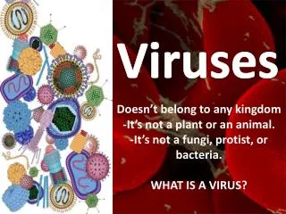
Evolution of Viruses Viruses do not fit our definition of living organisms Since viruses can replicate only within cells, they probably evolved after the first cells appeared Candidates for the source of viral genomes are plasmids (circular DNA in bacteria and yeasts) and transposons (small mobile DNA segments) Plasmids, transposons, and viruses are all mobile genetic elements