Download
1 / 22
230 likes | 343 Views
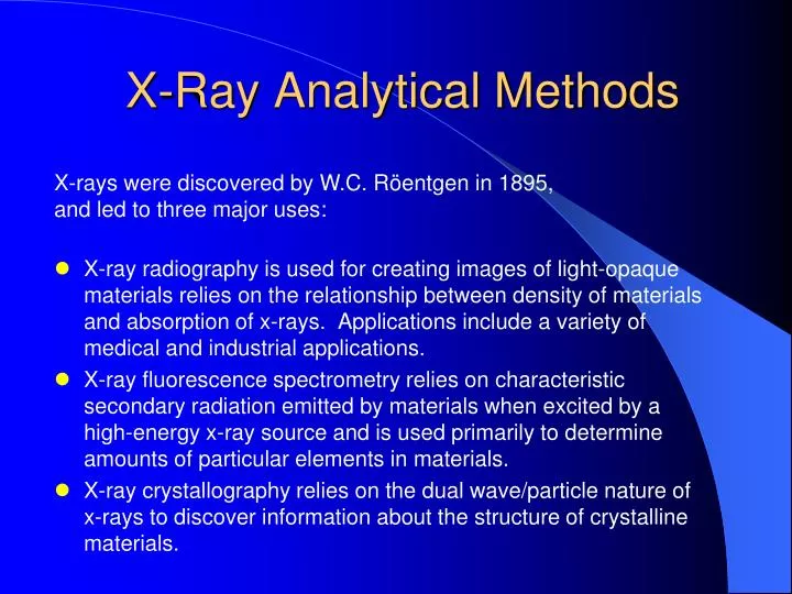
X-Ray Analytical Methods. X-rays were discovered by W.C. Röentgen in 1895, and led to three major uses:.

E N D
X-Ray Analytical Methods X-rays were discovered by W.C. Röentgen in 1895, and led to three major uses: • X-ray radiography is used for creating images of light-opaque materials relies on the relationship between density of materials and absorption of x-rays. Applications include a variety of medical and industrial applications. • X-ray fluorescence spectrometry relies on characteristic secondary radiation emitted by materials when excited by a high-energy x-ray source and is used primarily to determine amounts of particular elements in materials. • X-ray crystallography relies on the dual wave/particle nature of x-rays to discover information about the structure of crystalline materials.
Uses of X-Ray Powder Diffraction • Radiation safety for users of X-ray diffraction • The generation of X- rays • The geometry of diffraction • Bragg’s Law • Basics of X-ray crystallography • Sample and specimen preparation techniques • Basic error analysis for X-Ray diffraction data Basic material to be covered in this course:
Uses of X-Ray Powder Diffraction • Identification of single-phase materials – minerals, chemical compounds or other engineered materials. • Identification of multiple phases in microcrystalline mixtures (i.e., rocks) • Determination of crystal structure of identified materials • Identification and structural analysis of clay minerals (an introduction by Dr. Dewey Moore) • Recognition of amorphous materials in partially crystalline mixtures Analytical methods you should learn in this course:
Uses of X-Ray Powder Diffraction • Crystallographic structural analysis and unit-cell calculations for crystalline materials. • Quantitative determination of amounts of different phases in multi-phase mixtures by peak-ratio calculations. • Quantitative determination of phases by whole-pattern refinement. • Determination of crystallite size from analysis of peak broadening. • Determine of crystallite shape from study of peak symmetry. • Study of thermal expansion in crystal structures using in-situ heating stage equipment. Analytical methods that will be introduced in this course but not covered in detail :
XRD for Dummies: From Specimen to analyzed sample with minimal math Specimen vs. Sample – An important distinction Mathematics is unavoidable when dealing with x-rays and crystallography, but modern analytical methods, software and computers can eliminate much of the tedium of calculations Avoid “Black Box” errors by understanding what is going on in your analyses
The Bragg Equation where n is an integer is the wavelength of the x-raysd is the interplanar spacing in the specimen is the diffraction angle (discussed later!) The Bragg equation is the fundamental equation, valid only for monochromatic X-rays, that is used to calculate interplanar spacings used in XRD analysis.
Scintag Diffractometer HV Power Supply Spellman DF 3 Solid State High-voltage generator power supply
Scintag Diffractometer Detector Bicron Scintillation Detector and Monochromator
Scintag Diffractometer Detector Power Supply • From right to left: • Power module • Bias Power Supply • Signal Amplifier • Ratemeter (counter)
Determining Phases Present in a SpecimenThe “Legacy” Method • Determine d-spacings and intensities from experimental data (film or strip chart) • Use paper indexes to determine likely phases • Compare likely phases with database of standard minerals by matching peaks Problems with the method: Error prone,tedious and time consuming, Indexes difficult to search, particularly as database grows Multi-phase samples are very difficult to deal with
Determining Phases Present in a SpecimenThe “Modern” Method of Basic Phase Identification • Automated diffractometer is operated using electronic data acquisition and control software. • Data are collected in digital (not analog form). • Software is used to search, analyze, and determine the peak positions and intensities in the pattern. • Software is used to compare digital pattern with all (or a selected subset) of the ICDD “Powder Diffraction File” database of experimental and calculated patterns; software will “flag” possible phases present. • Identified patterns are visually compared with the sample data and best match(es) are selected.
Determining Phases Present in a SpecimenAdvanced Phase Identification Techniques Single phase samples can usually be identified easily using basic methods; multiphase samples require more work. • Identify dominant phase in pattern and digitally remove peaks associated with that phase. • Re-search the ICDD database using the “reduced” pattern. • Repeat process until all phases are identified.
Problems with Computerized Search/Match • Experimental patterns rarely match database patterns exactly (so pattern subtractions distort data for subsequent searches) • Preferred orientation effects • Crystal chemistry variation from ICDD database • Structural variations from ICDD database • Different materials with same structure can have same pattern • Peak shifts from systematic errors can throw automatic matches off • Axial-Divergence Error • Flat-Specimen Error • Specimen transparency (penetration) errors • Specimen displacement errors • Alignment errors • Spurious peaks from tube contamination Know your Sample; don’t accept unreasonable results!