Download
1 / 56
570 likes | 867 Views
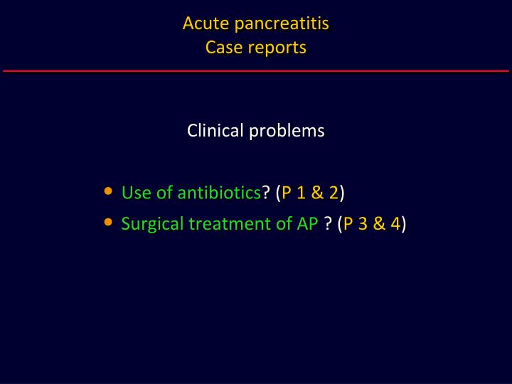
Acute pancreatitis Case reports. Clinical problems Use of antibiotics ? ( P 1 & 2 ) Surgical treatment of AP ? ( P 3 & 4 ). Acute pancreatitis Case reports. Case 1. Acute pancreatitis Case 1 – Patient KD. History M, 63 y Obesity – BMI 30.3 kg/m 2 Gallbladder stones

E N D
Acute pancreatitisCase reports Clinical problems • Use of antibiotics? (P 1 & 2) • Surgical treatment of AP ? (P 3 & 4)
AcutepancreatitisCasereports Case 1
Acute pancreatitisCase 1 – Patient KD History • M, 63 y • Obesity – BMI 30.3 kg/m2 • Gallbladder stones • No concomitant diseases • 1. episode of ABP • Time from onset 33.5 h
AcutepancreatitisCase 1 – Patient KD Lab data & prognosticassessment • WBC (G/l) 13.6 - 19.8 - 14.4 • CRP (mg/l) 57 - 167 - ND • RNS (pts) 5 • AP-O (pts 0-1-2) 9 - 8 - 8 • KCE (pts) 4
AcutepancreatitisCase 1 – Patient KD ERCP & CT • ERCP (day 0)CBD 5 mm, no stonesNo ES • CT (day 1)Mildinflammatoryinfiltrationclose to body and tail ofthepancreas and intheleftprerenalspace, no pancreaticnecrosis(BLT C, CTSI 2)
AcutepancreatitisCasereports Case 2
Acute pancreatitisCase 2 – Patient MK History • M,29 y • Overweight – BMI 28.5 kg/m2 • Gallbladder stones • No concomitant diseases • 1. episode of ABP • Time from onset 46.5 h
Acute pancreatitisCase 2 – Patient MK Lab data & prognostic assessment • WBC (G/l) 20.9 - 15.9 - 15.0 • CRP (mg/l) 71 - 95 - 166 • RNS (pts) 2 • AP-O (pts 0-1-2) 4 - 2 - 5 • KCE (pts) 2
AcutepancreatitisCase 2 – Patient MK ERCP & CT • ERCP (day 0)CBD 12 mm, impacted stone + 4 other stonesSE done, stones removed • TK (day 1)Moderate inflammatory infiltrations in both prerenalspaces, no pancreatic necrosis, small amount of fluid around the liver and mild bilateral hydrothorax(BLT C, CTSI 2)
Acute pancreatitisCases 1 & 2 • Case 1 - KD • Moderate prognosis • Case 2 - MK • Very good prognosis • SIRS (+) • Mild overweight / obesity • BLT C, no necrosis, CTSI 2 • WBC > 15 G/l • CRP > 150 mg/l Should antibiotics be administered?
Question • Whoshouldreceiveantibiotics? • Both • Patient 1 • Patient 2 • None
AcutepancreatitisCase 1 – Patient KD Bacteriology • Bile - positiveEscherichia coli(sensitive to Ciprofloxacin, Imipenem) • Blood – negative (1x)
AcutepancreatitisCase 2 – Patient MK Bacteriology • Blood – 10 x negative
AcutepancreatitisCase 1 & 2 • Wide-spectrumantibioticswereusedinbothcases • No complicationsacc. to Atlanta criteria • Probablymildnecrosis of peripancreaticfat • Hospitalstay 22 daysinbothcases • Coursemild / severe ?
AcutepancreatitisCasereports Case 3
Acute pancreatitisCase 3 – Patient AK History • M, 79 y • General condition severeHypertension 20 yParkinson’s disease? • Suspicion of gallstones • Probably 20 h from onset of abdominal pain • Very severe abdominal pain
AcutepancreatitisCase 3 – Patient AK OE • Dehydration • HR 116/min, RR 30/min • No peristalsis • Reboundtenderness+-
Acute pancreatitisCase 3 – Patient AK Angio-CT (day 0) • No mesenteric ischemia • Extensive atheromatosis • Extensive inflammatory infiltration of peripancreatic fat, non-enhancement area(up to 1/3) in body and tail • BLT C, necrosis < 1/3?, CTSI 4
AcutepancreatitisCase 3 – Patient AK Angio-CT(day 0)
Acute pancreatitisCase 3 – Patient AK ERCP (day 1) • Extensive swelling of D2 of moderate severity,bluish discoloration of mucosa, severe duodenopathy • Papilla very small and tight • No deep CBD cannulation despite pre-cut • CDB narrow (< 4 mm)
AcutepancreatitisCase 3 – Patient AK Lab data • WBC (G/l) 10.8- 20.1 - 15.3 • PLTS (G/l) 151 - 87 – 72 • HCT (%) 50 - 53 - 48 • paO2 (mm Hg) 101 - 65 - 52 • Cre (mg/dl) 1.1 - 0.9 - 1.6 • AT III (%) ND - ND – 42 • CRP ND!
AcutepancreatitisCase 3 – Patient AK Prognosticassessment • RNS (pts) 6 • AP-II (pts 0-1-2) 17 - 18 - 17 (deathrisk85%) • AP III J (pts 0-1-2) 50 - 65 - 57 (deathrisk67%) • KCE (pts) 7 • OFS (Bernard, pts 0-2) 1 - 6 (deathrisk85%)
AcutepancreatitisCase 3 – Patient AK CT (day 2) • Mildprogression • BLT E, necrosis< 1/3, CTSI 6
AcutepancreatitisCase 3 – Patient AK CT (day 2)
Acute pancreatitisCase 3 – Patient AK Clinical course • No improvement within 48 hours • Rapidly evolving multiorgan failure • Patient transferred to ICU • Surgical consultation
AcutepancreatitisCasereports Case 4
Acute pancreatitisCase 4 – Patient ML History • F, 50 y • No concomitant diseases • Mild obesity, BMI 31.6 kg/m2 • 10 months before single episode of biliary colicNo gallbladderstones • 1. episode of ABP • Time from onset 8 h
AcutepancreatitisCase 4 – Patient ML OE • Obesity • Jaundice • Epigastrictenderness
AcutepancreatitisCase 4 – Patient ML ERCP (day 0) • Duodenum and papillanormalCDB 10 mm, no stonesNo ESMicroscopic bile analysis: CMC+, CaBG+++
AcutepancreatitisCase 4 – Patient ML Lab data • WBC (G/l) 17.1 - 15.3 - 13.7 • HGB (g/dl) 14.3 - ND - 14.7 - 8.0 (d7) • paO2 (mm Hg) 73 • TP (mg/dl) 7.1 - 6.2 - 5.0 • CRP (mg/l)10- 68 - 200 - 298 (d7)
AcutepancreatitisCase 4 – Patient ML Prognosticassessment • RNS (pts) 6 • AP-O (pts 0-1-2) 8 - 9 - 6 • AP III J (pts 0-1-2) 23 - 22 - 19 • KCE (pts) 7 • OFS (Bernard, pts 0-2) 0 - 1
Acute pancreatitisCase 4 – Patient ML CT (day 2) • Enlarged pancreatic head, homogenous enhancement,no necrosis • Fluid collections at both prerenal spaces, in spleen hilum,between small bowel loops • BLT E, CTSI 4
Acute pancreatitisCase 4 – Patient ML Clinical course • Intensive conventional management, antibiotics • SIRS symptoms between days 10 and 16 • Control CT (d12) – progression, CTSI 4 • Control CT (d26) – progression, no pancreatic necrosis,but extensive necrosis of peripancreatic fat • Second period of fever from day 32, ↑ WBC i CRP • US – fluid collection, bacteriology – Str. faecalis • Surgical consultation (d43)
AcutepancreatitisCase 4 – Patient ML CT (days 2, 12 i 26)
AcutepancreatitisCase 4 – Patient ML CT (days 2, 12 i 26)
AcutepancreatitisCases 3 & 4 • Case 3 – AK • Day 3 • Sterilenecrosis • ↑ MOF • Bad prognosis Case 4 – ML • Day 43 • Infectednecrosis • No MOF • Moderateprognosis Whoshould be operated on?
Question • Whoshould be operated on? • Bothpatients • Patient 3 • Patient 4 • None
AcutepancreatitisCase 3 – Patient AK Surgery • Day 2 of hospitalization • 2000 ml brown fluid in the abdominal cavity • Extensive pancreatic necrosis (black pancreas)Necrosectomy. Setonage. Laparostomy • Cardiac arrest at the end of the procedure, death • Autopsy:Necrosis haemorrhagicapancreatis et telaeadiposae.Inflammatiopurulenta cum necrosi d. choledochi.
Acute pancreatitisCase 4 – Patient ML Surgery • Day 49 of hospitalization • Extensive fat necrosis • Abscess (500 ml) in lesser sac • Fat necrosis from right iliac fossa to diaphragmatic hiatus • Necrosectomy, setonage, laparostomy
Acute pancreatitisCase 4 – Patient ML Surgery (2) • Multiple exchanges of setones (11)days 51 to 68 • Wound abscess in the epigastrium6 drainage procedures from day 103 to 131 • Gradual improvement • Discharge on day 146
Acute pancreatitisIndications for surgery • Infectednecrosis • Localcomplications of pancreatitis • Sterilenecrosis
Duodenalswelling No swelling Minor swelling,limited to peripapillaryarea Moderateswelling withextensive involvementof D2 Severe swellingwith extensiveinvolvementof D2, bluishdiscoloration DGE MUSK 2000-2005
Duodenalswelling DGE MUSK 2000-2005
Duodenalswelling Normal duodenum Deformed duodenal loop D2 deformed and narrowed DGE MUSK 2000-2005
Duodenalswelling Edema of submucosal layer DGE & DPAT MUSK 2000-2005 Mucosal hyperemia