Download
1 / 43
460 likes | 1.18k Views
Towards safe practice in instrumental vaginal delivery. Leroy Edozien. Approximately 1 in 10 deliveries is instrumental. What could go wrong? Fetal complications. Subdural haematoma Subgaleal haematoma Cephalhaematoma Retinal haemorrhage Hyperbilirubinaemia. Facial laceration

E N D
Towards safe practice in instrumental vaginal delivery Leroy Edozien
What could go wrong?Fetal complications Subdural haematoma Subgaleal haematoma Cephalhaematoma Retinal haemorrhage Hyperbilirubinaemia Facial laceration Scalp laceration Facial nerve palsy Skull fracture Corneal injury Cervical spine injury
King SJ, Boothroyd AE. Cranial trauma following birth in term infants. Br J Radiol 1998;71:233-8
What could go wrong?Maternal complications Cervical laceration Haematoma Vaginal laceration Perineal tear Psychological trauma
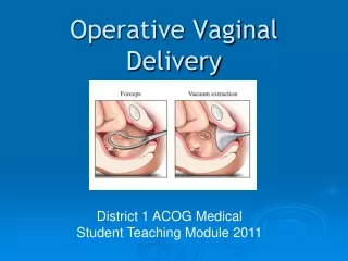
Avoiding harm Non-operative interventions Deciding when and when not to deliver instrumentally Using the right operative techniques
Non-operative interventions which reduce instrumental delivery rates • One-to-one support in labour (Hodnett, 2003) • Upright or lateral position (Gupta & Hofmeyr, 2003) • Oxytocin for prolonged second stage (Saunders et al, 1989) • Delayed pushing (Roberts et al, 2004)
When and when not to deliver instrumentally Indications: Fetal compromise (actual or anticipated) Prolonged second stage Where down-bearing is to be avoided
When and when not to deliver instrumentally Absolute contraindications: Malpresentation Unengaged fetal head Cephalopelvic disproportion Fetal clotting disorder GA < 34 wk (ventouse)
Safe practice: prerequisites for instrumental delivery • Fully dilated cervix • One-fifth or nil palpable abdominally • Ruptured membranes • Contractions present • Empty bladder • Presentation and position known • Satisfactory analgesia
Instrumental delivery before full cervical dilatation Crime or expedience? SOGC: ‘may be considered when benefits significantly outweigh risks’ RCOG: exceptions to the rule - cord prolapse at 9 cm in a multip; second twin
Engagement Instrumental delivery should not be attempted if the lowest part of the baby’s skull has not reached the ischial spines.
Smellie W. A treatise on the theory and practice of Midwifery. London; MDCCLII
Safe practice: abandonment Indications for abandonment: Difficulty in applying instrument No descent Delivery not imminent after three pulls 15 minutes elapsed
Why is the principle of abandonment frequently breached? Poor training Confirmation bias Sunk costs
Safe practice: recognise conditions predictive of difficulty/failure 1/5 palpable Station 0 OP position Moulding ++/+++ Slow progress Big baby BMI > 30 Trial of instrumental delivery
Sequential instrumentation Benefits and risks Decision-making
Safe practice: post-operative care Examine and observe the baby VTE risk assessment Bladder care Openness
Documentation Indication Abdominal examination Consent Position; station Moulding; caput Pelvis adequate CTG Contractions Ease of application No. of pulls Detachments Duration VE; PR post-delivery Condition of baby Cord pH Details of repair
Examples of error in instrumental delivery Action omitted, mistimed, misjudged: Abdominal palpation not done Prolonged traction Continuous traction Rotation during a contraction Traction directed forwards and upwards too soon
Examples of error in instrumental delivery Information wrong, incomplete or not retrieved: Mistaken head level or position Moulding not assessed Equipment not checked History of diabetes disregarded
Examples of error in instrumental delivery Procedural checks omitted or not properly done: No check for correct application No check for descent with pull PR/VE not done at end of procedure Swabs not counted
Examples of error in instrumental delivery Faulty selection (choosing from options): Wrong ventouse cup type Ill-advised sequential instrumentation
Examples of error in instrumental delivery Failure to communicate: With woman midwife senior obstetrician anaesthetist paediatrician
Examples of error in instrumental delivery Cognition: Failure to anticipate ….PPH, Shoulder dystocia, etc. Failure to ask the right questions e.g. pulling in the right direction? … forceps applied on baby’s face?
Training, competence supervision Unmet training needs Demonstrable benefits of training Assessment tools
‘Dr C stated that he discussed these options with Mr A and Mrs B and said that they were happy for him to deliver their baby using forceps. Mr A and Mrs B considered that Dr C did not communicate very effectively with them before or during the delivery. They said it was often very difficult to hear and understand what he was saying, particularly because Dr C directed most of his comments to Ms F.’
Assessment: occipito-posterior position, slightly to the right; presenting part slightly tilted.‘Dr C applied the left blade of the forceps directly to the baby’s head, followed by the right blade. As the handles could not be aligned properly he removed the blades and reassessed the position of the head. At this stage, Mrs B’s buttocks were brought down further towards the edge of the bed and Dr C removed the foetal scalp electrode to enable easier application of the forceps.Dr C explained that after re-examination he was satisfied that the baby was in an occipito-posterior position and so he reapplied the forceps. He stated that this time the blades aligned without difficulty. Dr C attempted to rotate the baby’s head to the right but was unable to and so attempted rotation to the left, which was also unsuccessful’
While kneeling on the floor, Dr C applied force on the forceps during a contraction, in an attempt to pull the baby down in the occipito-posterior position while Mrs B was asked to push. Dr C explained that sometimes the head can be rotated at a lower level, or delivered in that position without the need for any rotation. He stated that only moderate traction was applied during this procedure and that he only used his right forearm while his left arm was resting on top of his right hand.
Mr A and Mrs B stated that Dr C pulled extremely firmly on the forceps and that Mrs B was dragged down the bed as a result. Dr C denied using any more force than wasnecessary or than he would normally use during such a procedure.
‘Other than a small laceration on the left cheek of the baby from the scalpel blade atthe time of the operation, I did not see any external forceps marks or bruises on thebaby’s head or the face at the time of delivery’.-Dr C
Cord blood was obtained but had clotted and was unsuitable for pH analysis.
Baby born moribund. NICU. NND.This was Mrs B’s second pregnancy and the pregnancy had been uneventful. Her first child had died of a congenital heartdefect (at 20 weeks’ gestation).
Joint RCOG/ENTER MEETINGRisk Management and Medico-Legal Issues In Women’s Health25 to 26 April 2007