Download
1 / 41
430 likes | 670 Views
Proton Therapy . Marcio Fagundes, MD Radiation Oncology ProCure Oklahoma City Proton Center. Agenda. Why Protons Matter Differences between conventional x-ray (photon) radiotherapy and proton therapy How protons can provide a radiation dose advantage Clinical applications

E N D
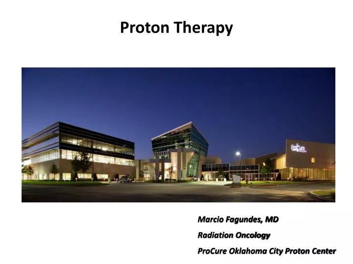
Proton Therapy Marcio Fagundes, MD Radiation Oncology ProCure Oklahoma City Proton Center
Agenda • Why Protons Matter • Differences between conventional x-ray (photon) radiotherapy and proton therapy • How protons can provide a radiation dose advantage • Clinical applications • Case illustrations
A Very Experienced Team in Proton Therapy: Development, Delivery and Operations Loma Linda UFPTI M.D. Anderson ProCure OKC Center MPRI MGH
The Value of Protons • Protons are physically superior to X-rays • Protons behave differently than x-rays: • Protons • X-Rays do not • Protons improve the “therapeutic ratio” • maximizing tumor control while minimizing side effects • At a given radiation dose to a tumor protons deliver, on average, less than half the radiation dose to normal tissues than do x-rays 1 • (1) Jay Loeffler, Massachusetts General Hospital, “Proton Therapy 2009”
Clinical applications for Proton Therapy • Head / Neck • Eye • Sinus/nasal • Throat • Ear • Pediatric • Brain • Spinal Cord • Bone • Neurologic • Brain • Spinal Cord • Other Solid Tumors • Breast Cancer • Lung Cancer • Colorectal Cancer • Prostate – select Source : National Cancer Institute
Indications for Proton Therapy “Classic” “Emerging” Lung Esophagus Whole pelvic RT Examples: Rectal & Anal Canal Large sarcomas Liver Mediastinal tumors Example: Lymphoma & Thymoma Extracranial radiosurgery Prostate and Lung SBRT • Pediatric tumors • Brain tumors • Base of skull tumors • Spinal tumors • Paraspinal tumors • Prostate cancers • Uveal melanomas • Intracranial radiosurgery
A different way to think about it • Tumors with: • curative intent or life expectancy beyond 2 years • in which anatomic motion is either minimal or can be accounted for • and there is something to avoid • Example: Pediatrics • Yes: Medulloblastoma • CSI avoids heart • Boost RT eliminates dose to supratentorial brain • No: Wilm’s Tumor • Target is often the entire peritoneal surface
The Goal of Radiation Therapy:Increase the Therapeutic Ratio • The goal of radiation oncology for 100 years has been to get: • More radiation in the tumor • Less radiation in the healthy tissue surrounding the tumor Tumor Control Normal Tissue Complications = Therapeutic Ratio
External-Beam Radiation Therapy • “EBRT is radiation from the outside-in” • This can be done in 2 ways: • X-Ray therapy (IMRT) • Proton therapy
X-ray (photon) radiotherapyIMRT (intensity modulated radiotherapy) Linear Accelarators Tomotherapy Cyber-knife
Evolution, Not Revolution Improvements in radiation dose distribution 6x Parallel opposed fields 3D 5 fields IMRT 9 fields Protons 4 field protons x-rays x-rays x-rays tumor
What is Proton Radiation?How is it different from X-rays and IMRT?
The Physics of Protons In order to deliver the same dose to the tumor, x-rays must deliver a greater dose outside the target than protons do Depth dose curves for protons and x-rays 300 Additional dose outside the target delivered with x-rays X-rays 250 200 Relative Dose (%) 150 100 50 Tumor Protons 0 0 50 100 150 200 250 300 350 400 Depth in Body (mm)
Protons Are More Precise and Spare Healthy Tissue 6 Field IMRT PLAN 3 Field Proton PLAN Tumor Tumor Exit dose unnecessarily radiates healthy tissues that may cause harmful side effects or secondary malignancies Zero exit dose and high degree of conformity eliminates excess radiation
Evidence of Distal Range Stopping Before treatment Treatment plan After treatment
Base of skull clivalchordoma-dose shown as percentages in the treatment plans-for physicians use IMRT-Protons: showing extra dose for IMRT IMRT Protons Dose volume histogram shown as actual doses
Base of skull clivalchordoma (Head)-anatomy Jaw Area to be treated Spinal cord Neck bone/vertebra Muscle
Increased TCP Increased dose No reductions Decreased NTCP Decreased dose to critical structures Kidneys Lungs Heart Blood vessels GI tract Why Protons for Paraspinal Sites? More dose and better tolerance of therapy
Paraspinal Ewing’s Sarcoma:A Case Comparison • A teenage football player ignoring a “right back bruise” for 4 months • Diagnosis: Paraspinal Ewing’s Sarcoma • Treatment per COG protocol: • 42 weeks of 5 drug chemotherapy • RT to primary site for local control
Diagnosis These are 2 different patients! Nearly Identical Location and GTV Patient on the left – 13 year old who presented in March 2006 Patient on the right – 16 year old who presented in June 2006
10% 50% Larger PTV needed for IMRT R Kidney
18 months post-RT Patient on the left – 13 year old who presented in March 2006 and was treated with X-Ray IMRT Patient on the right – 16 year old who presented in June 2006 who was treated with proton therapy
Why Protons for Lung Cancer? • X-rays have reached its dose limits • 3 trials showing that the maximum tolerated dose is 74 Gy with chemotherapy • RTOG 0117 Phase I • North Central Cancer Treatment Group • CALGB • Protons allow dose escalation while reducing toxicity compared to x-rays • Sejpal, M.D. Anderson 2011 • Chang, M.D. Anderson 2011 • Dose escalation can be achieved with protons without exceeding known indicators of lung toxicity • Chang, M.D. Anderson 2006 • For stage III tumors, radiation with chemotherapy holds the most promise • Clinical outcomes suggest better control rates and lower toxicity when using protons compared to x-rays with chemotherapy for stage III NSCLC • NCCN guidelines consider RT + chemo standard of care1 11http://www.nccn.com/images/patient-guidelines/pdf/nsclc.pdf 26
Lung/Mediastinum – IIIA NSCLC IMRT-Protons Protons IMRT Dose volume histogram shown as actual doses with TV to 70 CGE Dash – IMRT Solid – Proton Less dose to right (I/L) lung No dose to left lung Less dose to heart Less dose to spinal cord 27
Excess Radiation Causes Long-Term Side Effects • Radiation-induced pneumonitis can result from even low doses of excess radiation in the lungs (1,2) • Compared to IMRT, protons expose less lung tissue at all the critical/threshold doses listed below(5) which have been shown to be predictors of pneumonitis: • Mean dose to lung(1,2,3,4) • Volume of lung receiving 5 Gy(1,2,4) • Volume of lung receiving 10 Gy(2,4) • Volume of lung receiving 20 Gy(1,2,4) Comparison of Dose Escalated Proton Therapy and IMRT, both 74 Gy, for Stage III Lung Cancer • A Allen et al., “Fatal pneumonitis associated with IMRT radiation therapy for mesothelioma,” International Journal Radiation Oncology Biology Physics 65 (2006): 640-645 • E Yorke et al., “Correlation of dosimetric factors and radiation pneumonitis for non-small-cell lung cancer patients in a recently completed dose escalation study,” International Journal Radiation Oncology Biology Physics 63 (2005): 672-682 • Y Seppenwoolde et al., “Comparing different NTCP models that predict incidence of radiation pneumonitis,” International Journal Radiation Oncology Biology Physics 55 (2003): 725-735 • Liao et al., “Analysis of Clinical Dosimetric Factors Associated with Radiation Pneumonitis in Patients with Non-Small Cell Lung Cancer Treated with Concurrent Chemotherapy and Three Dimensional Conformal Radiotherapy,” International Journal Radiation Oncology Biology Physics 63 Supplement 1 (2005): S41 • J Chang et al., “Significant reduction of normal tissue dose by proton radiotherapy compared with three-dimensional conformal or intensity-modulated radiation therapy in stage I or stage III non-small-cell lung cancer,” International Journal Radiation Oncology Biology Physics 65 (2006): 1087-1096 28
M.D. Anderson lung toxicity data for inoperable locally advanced nsclc NSCLC treated with radiation therapy + chemotherapy1 1 Samir Sejpal et al., “Early findings on toxicity of proton beam therapy with concurrent chemotherapy for nonsmall cell lung cancer,” Cancer e-publication ahead of print (January 24, 2011): 1- 10 2 Retrospective analysis. In lieu of selection criteria, percentage of patients with Stage IIIA-IIIB were summarized
Summary of toxicity from other studies of x-ray radiation and chemotherapy Note: Please see Sejpal paper in Cancer 2011 for literature cited
Lung cancer lung trials Summary of lung trials for proton therapy 31 Source: clinicaltrials.gov search “protons AND lung AND radiation”
Therapy for Prostate Cancer Invasive Non-Invasive external-beamradiation therapy
Radiation Therapy (proton or IMRT)is an option after surgical failure radiation after surgery is easier to do than surgery after radiation
“Direct Radiation Complications Never Occur In Unirradiated Tissues” Dr. Herman Suit1 IMRT immerses more healthy tissue with radiation Radiation Therapy Plans for Prostate Cancer IMRT - 7-field co-planer Proton Therapy - 2-field DS Blue – 13% Green – 51% Purple – 63% Yellow – 76% Red – 95% Tumor Higher dose bath to healthy tissue with IMRT:Pelvis, rectum and bladder Less healthy tissue exposed to radiation compared to IMRT • Herman Suit, “The Grey Lecture 2001: Coming Technological Advances in Radiation Oncology,” International Journal of Radiation Oncology Biology Physics 53 No. 4 (2002): 798-809.
University of Florida Dosimetry Data Show Protons Reduce Dose To The Rectum By 59% IJROBP 2008 Radiation dose to the rectum – proton therapy and IMRT1 • Background on study • First prostate patients seen at University of Florida Proton Therapy Institute (“UFPTI”) • Both proton and IMRT plans were planned prospectively for each patient • The results • Relative and absolute mean rectal dose savings of 59.2% and 20.1%, respectively, with proton therapy • Why this is important • Entire Dose Volume Histogram (“DVH”) does matter, not just high the dose region • Rectal wall volume irradiated at 32.4 Gy is biggest predictor of rectal toxicity2 • Extremely high correlation between rectal volume irradiation to 70 Gy and 5-year toxicity rates3 90% 80% IMRT 70% 60% 50% Rectal Volume Receiving Radiation (%) Dose to rectum is more than 2x with IMRT vs. protons at 32 Gy 40% 30% Dose to rectum is almost 2x with IMRT vs. protons at 70 Gy 20% Proton 10% 0% 0 10 20 30 40 50 60 70 80 90 Radiation Dose (CGE/Gy) • Carlos Vargas et al., “Dose-Volume Comparison of Proton Therapy and Intensity-Modulated Radiotherapy for Prostate Cancer,” International Journal of Radiation Oncology Biology Physics 70 No.3 (2008): 744-751. • Susan Tucker, Lei Dong, Rex Cheung, et al., “Comparison of Rectal Dose-Wall Histogram Versus Dose-Volume Histogram for Modeling the Incidence of Late Rectal Bleeding After Radiotherapy,” International Journal of Radiation Oncology Biology Physics 60 (2004): 1589-1601. • Mark Storey, Alan Pollack, Gunar Zagars et al., “Complications from Radiotherapy Dose Escalation in Prostate Cancer: Preliminary Results of a Randomized Trial,” International Journal of Radiation Oncology Biology Physics 48 (2000): 635-642.
GI (Rectal) Side Effects and Complications The probability of damage to the GI tract is much higherwith x-rays than protons Chronic Radiation Proctitis in the GI tract Inflammation causedby radiation Necrosis and ulcer
Dose Escalation Trials Support the Use of Protons for Prostate Cancer Protons offer better control and lower toxicity than X-Rays The best outcome for control AND toxicity was achieved using protons • Alan Pollock et al., “Prostate cancer radiation dose response: results of M.D. Anderson Phase III randomized trial,” International Journal of Radiation Oncology Biology Physics 53 (2002): 1097-1105. (Note: toxicity updated from Viani et al, ref 6) • ST Peters, WD Heemsbergen, PC Koper et al., “Dose-response in radiotherapy for localized prostate cancer: results of the Dutch multicenter randomized phase III trial comparing 68 Gy of radiotherapy with 78 Gy,” 24 (2006): 1990-1196. • DP Dearnaley, MR Sydes, JD Graham et al, “Escalated-dose versus standard-dose conformal radiotherapy in prostate cancer: first results from the MRC RT101 randomized controlled trial,” Lancet Oncology 8 (2007): 475-487. • Anthony L. Zietman, “Correction: Inaccurate analysis and results in a Study of Radiation Therapy in Adenocarcinoma of the Prostate,” JAMA 299 No. 8 (2008): 898-900. Anthony L. Zietman et al., “Comparison of Conventional-Dose vs. High-Dose Conformal Radiation Therapy in Clinically Localized Adenocarcinoma of the Prostate. A Randomized Controlled Trial,” JAMA 299 No. 10 (2008): 899-900. • Veronique Beckendorf et al. “70 Gy vs. 80 Gy in localized prostate cancer: 5 year results of GETUG 06 randomized trial,” Int J Radiat Oncol Biol Phys (2011) epublication • Viani GA et al. Higher-than-conventional radiation doses in localized prostate cancer treatment: a meta-analysis of randomized, controlled trials. Int J Radiat Oncol Biol Phys. 2009 Aug 1;74(5):1405-18. • Note: Control rates are measured using the ASTRO definition, except for MRC RT01 which uses the Phoenix definition
Radiation-Related Coronary Artery Disease Atherosclerosis process similar to other CAD causes Intimal fibroblast proliferation macrophage plaque formation thrombus formation Ischemic heart disease Fig. 3. Left anterior descending coronary artery in a 16-year-old boy 1 year after receiving 40 Gy mantle radiotherapy for Hodgkin’s disease. Myointimal proliferation with considerable lumen narrowing. (Fajardo LF et al. ActaOncologica 44:13,2005)
U Penn • Review of 961 pts (1977-1994) • Median f/u 12 years • Survival free from • Coronary Artery Disease (CAD) • 90% Right-sided • 75% Left-sided “Significantly higher rate of fatal and non-fatal diagnosis of coronary artery disease seen in left-sided patients compared with right-sided XRT” Harris, E et al. JCO 24:4100,2006
Coronary Artery CT Scan Distal LAD 2nd Diagonal
Protons and Electron/X-Ray Match Protons Electron / X-Ray Match Difference