Download
1 / 42
440 likes | 622 Views
Introduction Spiral Cone-beam CT Interior Tomography Multi-scale CT Work in Progress. Outline. Integrated Biomedical Research Program 3 Medical Schools 1 Vet-Med College 10 Institutes/Centers 51 Faculty (17 Primary). Virginia Tech. Established in 1872

E N D
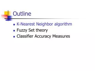
Introduction Spiral Cone-beam CT Interior Tomography Multi-scale CT Work in Progress Outline
Integrated Biomedical Research Program • 3 Medical Schools • 1 Vet-Med College • 10 Institutes/Centers • 51 Faculty (17 Primary)
Virginia Tech • Established in 1872 • Ranked 25th for College of Engineering • Ranked 12th in Patents • Ranked 8th in Licenses to Start-ups • Best multi-scale CT facility
Wake Forest University • Medical School Established in 1902 • Ranked 30th among National Universities • Ranked 32th in NIH Funding • Ranked 1st in Regenerative Medicine (WFIRM)
Biomedical Imaging Bioluminescence & Fluorescence Nuclear Imaging CT/X-Ray MRI
Bioluminescence Tomography (2002) Computed Tomography Individualized Volume Shape Model Optical Tomography Optical Model Bioluminescence Imaging 3D Mapping Bioluminescent Views Wang G, et al.: In vivo mouse studies with bioluminescence tomography. Optics Express 14:7801-7809, 2006
Introduction Spiral Cone-beam CT Interior Tomography Multi-scale CT Work in Progress Outline
Scanning for Faster Speed Ray Sum Spiral into Modern CT Era
Interpolation to Planar Data z z y x 1D Detector Array Ray A
Spiral Cone-beam CT (1991) Wang, G, Lin, TH, Cheng PC, Shinozaki DM, Kim, HG: Scanning cone-beam reconstruction algorithms for x-ray microtomography.Proc. SPIE Vol. 1556, p. 99-112, July 1991 (Scanning Microscopy Instrumentation, Gordon S. Kino; Ed.)
Multiple Implications To solve the long-object problem, a first level of improvement with respect to the 2D FBP algorithms was obtained by backprojecting the data in 3D, along the actual measurement rays.The prototype of this approach isthe algorithm of Wang et al. Defrise, Noo, Kudo: A solution to the long-object problem in helical cone-beam tomography. Phys. Med. Biol. 45:623-643, 2000 Many advances in CB reconstruction have been made recently thanks to the quest for an attractive reconstruction method in helical CB tomography. Pack, Noo, Clackdoyle: Cone-beam reconstruction using the backprojection of locally filtered projections. IEEE Trans. Medical Imaging 24:1-16, 2005
Seeking Exact Solution (1997) “Cone-beam spiral CT seems an ideal imaging mode. It is desirable and possible that an exact cone-beam reconstruction algorithm be designed that takes longitudinally truncated cone-beam data and is computationally efficient.” Wang G, Cheng PC, Vannier MW: Spiral CT - Current status and future directions. Proc. SPIE 3149 203–12, 1997
Katsevich Formula (2002) Object Source Pi-Line Detector Plate Katsevich A: A general scheme for constructing inversion algorithms for cone beam CT. Int'l J. of Math. and Math. Sci. 21:1305-1321, 2003
Annually, ~100M CT scans are performed in USA alone h-index=30
Dual-source CT (2003) Bioluminescence tomography prototype Designed by Wang, Hoffman, McLennan Built by us & UI Med. Inst. Facility in 2003 Dual-source In vivo Micro-CT scanner Designed by BIR, Hoffman, Wang Built by BIR in 2003
Introduction Spiral Cone-beam CT Interior Tomography Multi-scale CT Work in Progress Outline
Inner Vision with Local Data y t x Measurement Projection:Linear integrals Sinogram X X Object t X-rays Reconstruction
Extension to Other Modalities GeWang, HengyongYu
Introduction Spiral Cone-beam CT Interior Tomography Multi-scale CT Work in Progress Outline
Nano-CT Momentum Andrew G. Peele et al.: High Resolution X-ray phase tomography. Proc. SPIE CT Conference, 2010
X-ray Beam Central Stop Sample Phase Ring Zone Plate Detector Plane Condense Lens ROI Sample Stage Interior Nano-CT
Potential for Study on Earliest Life Hagadorn JW, et al. (2006) Cellular and subcellular structure of Neoproterozoic embryos. Science 314:291–294
Introduction Spiral Cone-beam CT Interior Tomography Multi-scale CT Work in Progress Outline
Dark-field Tomography Wang G, Cong W, Shen H, Zou Y: Varying Collimation for Dark-Field Extraction. International Journal of Biomedical Imaging. 2009, Article ID 847537, 2010
Important collaborators include, but not limited to, Drs. E. W. Bai, J. A. Brink, Z. Q. Chen, P. C. Cheng, W. X. Cong, M. Furth, W. M. Han, M. Jiang, A. Katsevich, Y. Li, L. Li, H. O. Shen, D. M. Shinozaki, D. L. Snyder, S. Soker, M. W. Vannier, S. Wang, Y. B. Wen, C. Wyatt, Y. Xu, J. S. Yang, Y. B. Ye, H. Y. Yu, J. Zhao, T. G. Zhuang, and Y Zou. This work is partially supported by multiple NSF, NIH and industrial grants. Acknowledgement