Download
1 / 24
260 likes | 528 Views
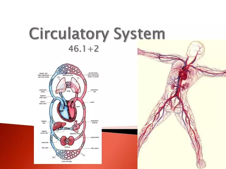
Circulatory System. 46.1+2. 46.1. Cardiovascular system = the blood, heart, & blood vessels Lymphatic system = the lymph, lymph nodes, & lymph vessels Together they make the circulatory system. Functions of the Circulatory System. Transports nutrients, hormones, & gases

E N D
Circulatory System 46.1+2
46.1 • Cardiovascular system = the blood, heart, & blood vessels • Lymphatic system = the lymph, lymph nodes, & lymph vessels • Together they make the circulatory system
Functions of the Circulatory System • Transports nutrients, hormones, & gases • Get rid of wastes • Helps maintain a constant body temperature http://www.youtube.com/watch?v=H04d3rJCLCE
Superior Vena Cava Inferior Vena Cava Aorta Left Ventricle Right Ventricle Pulmonary Veins (twice) Pulmonary Artery Right Atrium Left Atrium Tricuspid Valve Semilunar Valves Mitral Valve Septum RIGHT LUNG LEFT LUNG
Your Heart • Main function of the heart is to keep blood moving constantly throughout the body. • Large organ made of cardiac muscle that are rich with mitochondria. Surrounded by a tough membrane called the pericardium. • Mammalian hearts have four chambers. • atria • Ventricles Left and right side of the heart are separated by the septum.
Parts of the Heart • Aorta: large blood vessel • Vena cava: Sends deoxygenated blood into the right atrium (Superior from upper body; Inferior from lower body) • Atria: Two upper chambers • Walls thinner and less muscular than ventricles • Ventricles: Two lower chambers • Walls thicker and more muscular than atria (why?) • Left pumps blood to entire body; thicker than right ventricle (why?) • Right pumps blood to the lungs
1) Why is the heart’s left side exerting more pressure? • 2) Why is the left side’s wall of muscle larger than the right side’s?
Heartbeat Control • Sinoatrial Node (SA Node)-Initiates an electrical impulse to contract the heart. • located in the right atrium. • Also called the ‘pacemaker’ for the heart. • Impulse travels next to the atrioventricular node (AV Node) • Located in the septum • Causes the ventricles to contract. • Muscle cells contract in waves http://www.youtube.com/watch?v=waOSUpEHPQs&feature=related
Pacemaker Implantation http://www.youtube.com/watch?v=-cDSVytVGhc
Blood Pressure: • systolic pressure- when the ventricles contract • diastolic pressure- when the ventricles are relaxed • Pulse: Pressure waves in arteries from contraction of the left ventricle Hypertension- high blood pressure http://www.youtube.com/watch?v=Nb4jpp-GGUs
SYSTOLE: CONTRACT DIASTOLE: RELAX
Blood Vessels-Arteries and Veins • Arteries: large thick walled muscular elastic vessels that carry oxygenated blood away from the heart. • Blood is under pressure pushed through by heart pumping. (blood pressure) • Arteries branch off from heart divide into smaller vessels called arterioles. • Arterioles enter tissues where they branch in capillaries. • Veins: large blood vessels that carry deoxygenated blood from tissues back toward the heart. • Blood is not under pressure. • Veins in arms and legs have valves that prevent blood from flowing backward. • Muscles used to move blood • Several venules will merge to form veins. http://www.youtube.com/watch?v=CjNKbL_-cwA
Capillaries • Microscopic blood vessels. • Walls are one celled thick • Blood cells move through in single file line. • Capillary walls enable nutrients and gases to diffuse easily between blood cells and surrounding tissue cells. http://www.youtube.com/watch?v=NF68qhyfcoM&feature=related
Artery, Vein, Capillary http://www.wisc-online.com/Objects/ViewObject.aspx?ID=AP12704
FUN FACT What color is blood inside the body? If it is red then why do veins look blue? Blood is a bright red in its oxygenated form (i.e., leaving the lungs), when hemoglobin is bound to oxygen to form oxyhemoglobin. It's a dark red in its deoxygenated form (i.e., returning to the lungs), when hemoglobin is bound to carbon dioxide to form carboxyhemoglobin. Veins appear blue because light, penetrating the skin, is absorbed and reflected back to the eye. Since only the higher energy wavelengths can do this (lower energy wavelengths just don't have the *oomph*), only higher energy wavelengths are seen. And higher energy wavelengths are what we call "blue." In an experiment, glass tubes were filled with blood and immersed in milk, milk having a similar ratio of fat, proteins, and water in emulsion as skin. At a certain depth, the tubes appeared blue. A phlebotomist (a person who draws blood) from the local university hospital drew hundreds of samples of venous blood. They said that almost all samples were dark red. Those that weren't were usually because people were on enriched oxygen systems. At the same time, respiratory therapists drew blood from an artery, not a vein. These samples were characteristically bright red, unless the patient was having difficulty breathing (asthma, people with one lung, pneumonia, emphysema, whatever). DEOXYGENATED BLOOD -DEEP RED OXYGENATED BLOOD -BRIGHT RED
Patterns of Circulation • Pulmonary Circulation- blood traveling between the heart and lungs. -pulmonary veins and arteries • Systemic Circulation- blood traveling between the heart and other body tissues. • coronary circulation (heart) • atherosclerosis = build-up of fatty material on the inside of coronary arteries • renal circulation (Kidney) • hepatic portal circulation (Liver) http://www.youtube.com/watch?v=xU30BltUa4g&feature=plcp&context=C3d90551UDOEgsToPDskL-xtnUbfcc49ivlLNr8nht
Lymphatic System • Functions in returning fluids that have collected in the tissues to the bloodstream. • Fluid is called lymph. • No “pump” for the lymphatic system • Uses skeletal muscles • Valves prevent backflow • Lymph nodes filter lymph • Trap foreign particles, microorganisms, and tissue debris • Store lymphocytes, type of white blood cell that are specialized to fight disease and infection. • Swollen lymph nodes=increase in lymphocytes
Blood (46.2) • Plasma- liquid part of the blood, mostly water. Carries nourishment for the cells. • Red Blood Cells (erthrocytes)- transport oxygen. Formed in the red marrow of cells. -Hemoglobin- transport oxygen and carbon dioxide. -no nuclei -life span of 120-130 days
White Blood Cells (leukocytes)- help defend the body against disease. -formed in the red marrow, lymph nodes, and spleen. -larger than RBC’s -can live many years -several types ex: phagocyte http://www.youtube.com/watch?v=KxTYyNEbVU4
Platelets- help form blood clots. • Fibrin (protein) is produced that forms a sticky web, forming a clot. • Hemophilia-absence of one or more proteins needed to clot • Blood Types- A, B, O, AB • Antigens- protein or carbohydrate that signals the body that something foreign has entered it. • Rhfactor- antigen present on the surface of RBC’s. Majority of humans are Rh +.