Download
1 / 65
650 likes | 714 Views
Section H Protein synthesis. The genetic code Translation in prokaryotes Translation in eukaryotes. H 1 The genetic code. 1. Amino acids in a polypeptide chain were found to be coded by groups of three nucleotides in a mRNA.

E N D
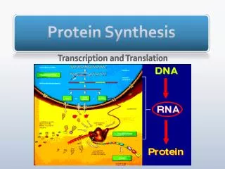
Section HProtein synthesis • The genetic code • Translation in prokaryotes • Translation in eukaryotes
1. Amino acids in a polypeptide chain were found to be coded by groups of three nucleotides in a mRNA • Simplecalculation indicated that three or more bases are probably needed to specify one amino acid. • Genetic studies of insertion, deletion, and substitution mutants showed codons for amino acids are triplet of nucleotides; codons do not overlap and there is no punctuation between codons for successive amino acid residues. • The amino acid sequence of a polypeptide is defined by a linear sequence of contiguous codons: the first codon establishs a reading frame.
Altered amino acid sequences Genetic studies showed that genetic codons are successive triplets of nucleotides
Each mRNA molecule would have three potential reading frames (but only one usually codes for a polypeptide chain) .
2. All 64 triplet codes were deciphered by 1966 • 61 of the codons code for the 20 amino acids and three (UAA, UAG, UGA) for chain termination, called termination codons, stop codons, or nonsense codons). • AUG is a dual codon coding for initiation and Met. • 18 of the amino acids are coded by more than one codon: the genetic codes are degenerate (兼并性). (Having more than one codon specify the same amino acid.) • Condons that specify the same amina acid (synonyms) often differ only in the third base, the wobble(变偶or 摆动)position, where base-pairing with the anticodon may be less stringent than for the first two positions of the condon.
The codes seem to have evolved in such a way to minimize the deleterious effects of mutations, especially at the third bases: XYU and XYC always encode the same amino acid XYA and XYG usually code for the same amino acid. • A reading frame codes for more than 50 amino acids without a stop codon is called an open reading frame( (ORF), which has the potential of encoding a protein.
All 64 genetic codes
Established the chemical structure of tRNA Established the in vitro system for revealing the genetic codes Devised methods to synthesize RNAs with defined sequences
3. The genetic code has been proved to be nearly(not absolutely) universal • Direct comparisons of the amino acid sequences of proteins with the corresponding base sequence of their genes or mRNAs, as well as recombinant DNA technologies, proved that the genetic codes deciphered from in vitro studies were correct and almost universally applicable. • A small number of “unusual codes” have been revealed in many mitochondria genomes and nuclear genome of a few organisms.
4. Overlapping genes were found in some viral DNAs Next summary
遗传密码的基本性质: • 方向性 • 兼并性 • 通用性与例外 • 读码的连续性 • 变偶性 • 起始密码和终止密码
1. Three kinds of RNA molecules perform different but cooperative functions in protein synthesis • mRNAscarry the genetic information copied from DNA in the form of genetic codons. • tRNAs mediate the incorporation of specific amino acids according to genetic codons present on the mRNA molecules via their specific anticodon triplets. • rRNAs associate with a set of proteins to form the protein-synthesizing machines (ribosomes) and probably catalyze peptide bond formation during protein synthesis.
2. All tRNAs have common structural features:cloverleaf in secondary, “L” in 3-D structures.
3. Some tRNA molecules can recognize more than one codons via wobble pairing • The adapter tRNAs recognize the codons on a mRNA via a triplet called anticodons. • It was first proposed that a specific tRNA anticodon would exist for every of the 61 (or 64) codons, but less tRNAs were revealed. • It was revealed that highly purified tRNA molecules (e.g., alanyl-tRNAAla) of known sequence could recognize several different codons. • Inosine, which may form base pair with A, U, and C, was found to be present at the first position of the anticodons in some tRNAs.
Crick proposed the “wobble hypothesis” in 1966 to explain the pairing features between anticodons and codons: • The first two bases of a codon in mRNA confer most of the coding specificity, the third base can be loosely paired with the anticodons; • The firstbase of some anticodons can wobble and determines the number of codons a given tRNA can read (A and C for one, U and G for two, I for three); • Codons that specify the same amino acid but differ in either of the first two bases need different tRNAs, i.e., a mininum of 31 tRNA are needed to translate the 61 codons;
This hypothesis has been widely supported by all the evidence gathered since (thus the “wobble rule”). • This moderate pairing strength may serve to optimize both the accuracy and speed of polypeptide synthesis.
The codon-anticodon pairing between the a mRNA and a tRNA: the presence of an inosinate residue at position one in the anticodon allows the tRNA to recognize a few codons.
U G I C Possible wobble pairing between anticodon and codon. U A I I
4. The synthesis of a protein can be divided into five stages • Each amino acid is first covalently attached to a specific tRNA molecule in a reaction catalyzed by a specific aminoacyl-tRNA synthetase (Stage 1). • The mRNA then binds to the smaller subunit of the ribosome, after which the initiating aminoacyl-tRNA and the large subunits of the ribosome will bind in turn to form the initiating complex (Stage 2)
The first peptide bond is then formed after the second aminoacyl-tRNA is recruited with help of the elongation factors, and the chain is then further elongated (stage 3). • When a stop codon (UAA, UAG, and UGA) is met, the extension of the polypeptide chain will come to a stop and is released from the ribosome with help from release factors (Stage 4). • The newly synthesized polypeptide chain has to be folded and modified (in many cases) before becoming a functional protein (Stage 5).
5. The 20 aminoacyl-tRNA synthetases attach the 20 amino acids to one or more specific tRNAs (stay 1) • An amino acid is first activated to form an aminoacyl-AMP intermediate, and is then charged to one or more specific tRNAs all catalyzed by one such specific aminoacyl-tRNA synthetase.
Aminoacyl-tRNA synthetases can be divided into two classes based on differences in structure and reaction mechanisms.
(1)Amino acid + ATP aminoacyl-AMP +PPi (2)Aminoacyl-AMP + tRNA aminoacyl-tRNA + AMP The overall reaction is Amino acid + ATP + tRNA aminoacyl-tRNA + AMP +PPi
Aminoacyl-tRNA synthetase 3 binding sites in the active site Aminoacyl- tRNA
Stage 1 Each amino acid is specifically attached to a specific tRNA before used for protein synthesis
6. Translation in prokaryotes begins by the formation of a complex. (Stage 2 Initiation of protein synthesis) The Shine-Dalgarno sequence and a nearby AUG marks the start site of translation on a bacterial mRNA.
A purine-rich Shine-Dalgarno sequence and a AUG codon marks the start site of polypeptide synthesis on bacterial mRNA molecules.
This purine-rich sequence was found to be complementary to a pyrimidine-rich sequence at the 3`end of the 16S rRNA present in the 30S subunit of the ribosomes. • The Shine-Dalgarno sequence may thus serve as the ribosome binding site on bacterial mRNAs.
Stage 2 The initiating complex is assembled from the small subunit of the ribosome, the mRNA, the initiating aminoacyl-tRNA (being fMet-tRNAfMet in bacteria), and the large subunit of the ribosome.
The Met charged on tRNAiMet (by Met-tRNA synthetase) is specifically formylated by a transformylase to form fMet-tRNAfMet in bacteria (Met-tRNAMet can not be formylated). • fMet-tRNAfMet can only enter and only fMet-tRNAfMet can enter the initiation site (site P) on the ribosome in E.coli.
The initiating complex is assembled from the small subunit of the ribosome, the mRNA, the initiating aminoacyl-tRNA (being fMet-tRNAfMet in bacteria), and the large subunit of the ribosome.
Each prokaryotic ribosome has three binding sites for tRNA. aminoacyl-tRNA binding site A site peptidyl-tRNA binding site P site exit site E site initiation factors IFs N-formylmethionine fMetf
7. The polypeptide chain is elongated via a repeating three-step reactions(stage3) The elongation cycle consists of three steps aminoacyl-tRNA binding peptide bond formation translocation
The next aminoacyl-tRNA is delivered to the A (aminoacyl) site of the ribosome by EF-Tu-GTP (step 1). • The fMet group is transferred to the second aminoacyl-tRNA in the A site to form a peptide bond, generating a dipeptidyl-tRNA (step 2). • The 23S rRNA of the large subunit of the ribosome seems to have the peptidyl transferase activity, thus being a ribozyme (Noller, 1992).
EF-G (the translocase) then promotes the translocation of the ribosome along the mRNA by the distance of one codon: the deacylated tRNAfMet is released from the E (exit) site of the ribosome, the dipeptidyl-tRNA is relocated to the P site, and the A site is open for the incoming (the third) aminoacyl-tRNA (step 3).
Step 2: peptide bond formation (23S rRNA) Stage 4 Step 3: Translocation (EF-G) Step 1: Aminoacyl- tRNA enters the A site (EF-Tu) AA
The EF-Tu-GTP- aminoacyl-tRNA complex EF-Tu EF-G has a structure similar to EF-Tu-tRNA Aminoacyl-tRNA
8. Polypeptide synthesis is terminated by release factors that read the stop codons • Two bacterial protein factors called RF1 and RF2 recognize the stop codons (RF1 for UAA and UAG; RF2 for UAA and UGA). • A third releasing factor RF3 is a GTP-binding protein and seems to act in concert to promote the cleavage of the peptide from peptidyl-tRNA. • The specificity of the peptidyl transferase (23S rRNA) is altered by the release factor: water become the acceptor of the activated peptidyl moiety.