Download
1 / 36
500 likes | 665 Views
Uterine Myoma. Women’s Hospital, School of Medicine Zhejiang University Prof. Lin Jun. Introduction. Most common benign tumor of female reproductive system Benign neoplasm composed primarily of smooth muscle and connectives tissue Common in women 30-50y Usually asymptomatic.

E N D
Uterine Myoma Women’s Hospital, School of Medicine Zhejiang University Prof. Lin Jun
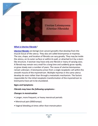
Introduction • Most common benign tumor of female reproductive system • Benign neoplasm composed primarily of smooth muscle and connectives tissue • Common in women 30-50y • Usually asymptomatic
Etiology Not detectable before puberty and after menopause Probably relates to female hormones • Estrogen and ER ↑ • Progestin may promote mitosisof myoma
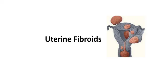
Classification According to the location: • Uterine body myoma 90% • Cervical myoma 10%
Classification According to the relationship between myoma and uterine myometrium : • intramural myoma60%-70% • subserous myoma20% • submucous myoma 10%-15%
Classification multiple myoma
Pathology Gross Appearance: • round, smooth, and usually firm • false capsular covering ——pseudocapsule can be clearly demarcated from the surrounding myometrium
Pathology Gross Appearance: Transverse section : • light gray • a whorl-like arrangement or an intertwining pattern
Pathology Microscopic examination: • composed of smooth muscle cells and varying amounts of connectives tissue • Individual cells are quite uniform in size, spindle shaped, have elongated nuclei. • Nonstriated muscle fibersare arranged in interlacing bundles of varying size running in different directions.
Degeneration • Hyaline degeneration • Cystic degeneration • Red degeneration • Sarcomatous change • Degeneration with calcification
Degeneration Red degeneration • most common during pregnancy and puerperium • venous thrombosis and congestion with interstitial hemorrhage
Degeneration ← Red degeneration ← Hyaline degeneration
Degeneration Sarcomatous change • malignant • rare, 0.4-0.8% • old women • enlarge rapidly with irregular vaginal bleeding
Degeneration Sarcomatous change
Symptoms • Usually no symptoms • Associate with location, and degenerations • Not associate with the size and the number
Symptoms 1. menorrhagia and prolonged menses • large intramural myoma • submucous myoma 2. abdominal mass 3. leukorrhagia
Symptoms 4. pressure effects • pressure bladder or rectum → urinary frequency, constipation • intraligamentous myomaand large cervical myoma → obstruct ureter 5.others • infertility • spontaneous abortion • abdominal pain
Sign associated with: • size • location • number • degeneration large myoma→ palpable abdominal mass Pelvic examination: uterus —— enlarged,irregular and hard
Diagnosis • Typical symptoms and signs • Ultrasound • Hysteroscopy • Laparoscopy
Diagnosis Hysteroscopy
Diagnosis Laparoscopy
Differentialdiagnosis • Pregnancy • Ovarian neoplasms • Adenomyosis • Malignant tumors of uterus • uterine sarcoma • endometrial carcinoma • cervical cancer
Treatment According to : • age • desire for childbearing • symptoms • location , size and amount of myoma
Treatment Observation and Follow Up • Small,asymptomatic,especially near menopause • Interval:3~6 months
Medical measure Indications: • smaller than 2 months in size • slight symptoms • near menopause
Medical measure 1.Androgenic agents: testosterone propionate 2.Gonadotropin-releasing hormone agonist, (GnRH-a) GnRH-a LH、FSH↓ E2↓ shrinkage of myoma • leuprorilin • goserelin
Medical measure • 2.GnRH-a • Side effects: • Hypoestrogenic side effects • Osteoporosis 3.Mifepristone
Surgical measures Indications: • greater than 10 weeks in size • menorrhagia→ anemia • pressure effects • grows rapidly • failure in medical treatment • infertility or recurrent abortion
Surgical measures Approaches: • laparotomy • hystereoscopy • laparoscopy
Surgical measures • Myomectomy • preserve fertility, <35 years old 2. Hysterectomy • Large myoma • Numerous tumors • Obviously symptomatic patient • No wish of preserving fertility • Suspected to malignant transformation
Myomas during pregnancy Impact on pregnancy and delivery : • abortion • preterm labor • fetal malpresentation • placenta previa • birth canal obstruction • postpartum hemorrhage
Myomas during pregnancy Red degeneration • Clinical finding: • rapid growth of myoma • pain, fever, WBC↑ • Conservative treatment