Download
1 / 47
550 likes | 1.13k Views
Intracellular Control of Cell-Cycle. First At the replication of DNA during S phase Second At the chromosome segregation and cell division of M phase. In yeast. S-phase cyclin-Cdk complexes ( S- Cdks ) initiate DNA replication once per cycle

E N D
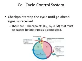
Intracellular Control of Cell-Cycle First At the replication of DNA during S phase Second At the chromosome segregation and cell division of M phase.
In yeast • S-phase cyclin-Cdk complexes (S-Cdks) initiate DNA replication once per cycle • DNA replication begins at origins of replication, which are scattered at various locations in the chromosome • Origin recognition complex (ORC) is a multiprotein complex that binds to the yeast replication origin • ORC bind to replication origins throughout the cell cycle and serve as landing pads for several additional regulatory proteins
The assembly of the pre-replicative complex (pre-RC) • In late mitosis and early G1, the proteins cdc6 and cdt1 bind to the ORC at origins • This leads to the binding of the Mcm proteinscomplex which is composed of a group of 6 closely related proteins • The resulting large complex is the pre-RC, and the origin is now licensed for replication.
The activation of S-Cdk in late G1 initiates DNA replication: • S-cdkphosphorylates Cdc6 which is then degraded • S-cdk trigger the assembly preinitiation complex • S-cdk with help of additional kinase collaborate to phosphorylate ORC. • DNA synthesis begins .
Several cyclin-Cdkcomplexes cooperate to restrain pre-RC assembly and prevent DNA rereplication after S phase • S-Cdk • It phosphorylatesCdc6 • It causes the Cdc6 protein to dissociate from ORC after an origin has fired • thus triggering its ubiquitylation by the SCF enzyme complex and thus its degradation • It also phosphorylates excess Mcm proteins • the Mcm protein complex cannot bind to a replication origin • this triggers their export from the nucleus
several cyclin-Cdk complexes cooperate to restrain pre-RC assembly and prevent DNA rereplication after S phase • This results in: • the disassembly of the pre-RC, which prevents replication from occurring again at the same origin • Preventing the Cdc6 and Mcm proteins from reassembling at any origin • S-Cdk activity remains high during G2 and early mitosis • preventing rereplication from occurring after the completion of S phase.
several cyclin-Cdk complexes cooperate to restrain pre-RC assembly and prevent DNA rereplication after S phase • M-Cdk • helps ensure that rereplication does not occur during mitosis by: • phosphorylating the Cdc6 and Mcm proteins. • G1/S-Cdks • Ensures that excess Mcm proteins unbound to origins in late G1 are taken out of action before replication begins by: • inducing Mcm export from the nucleus,
M-Phase Cyclin-Cdk Complexes (M-Cdks) Trigger Entry into Mitosis • After S phase the G2 cell is left with two sister chromatids glued together along their length. • In M phase the duplicated chromosomes and other cell contents are distributed equally to the two daughter cells. • The events of mitosis are triggered by M-Cdk, which is activated after S phase is complete.
After S phase is complete Increase in M-cyclin gene transcription during G2 and Mdecreased degradation of M-cyclin • Polo kinase, phosphorylates and partialactivates Cdc25 leads topartial activation of a subpopulation of M-Cdk complexes Accumulation of M-cyclin (cyclin B in vertebrate cells) The crucial event in late G2 • Positive feedbackphosphorylates and activates Cdc25 • Positive feedback Phosphorylates and inhibits Wee1.
What, then, triggers the activation of the M-Cdk stockpile? • The positive feedback loop converts a gradual increase in M-cyclin levels into a switch-like, abrupt rise in M-Cdk activity. • Similar molecular switches operate at various points in the cell cycle to ensure that events such as entry into mitosis occur in an all-or-none fashion.
Entry into Mitosis Is Blocked by Incomplete DNA Replication:The DNA Replication Checkpoint • Ensures that the initiation of mitosis cannot occur until the last nucleotide in the genome has been copied • No broken or incomplete sets of chromosomes are passed to the daughter cells. • Sensor mechanisms, of unknown molecular nature, detect either: • the unreplicated DNA • or the corresponding unfinished replication forks • It send a negative signal to the cell-cycle control system, blocking the activation of M-Cdk.
Protein kinase • If DNA is unreplicated or the corresponding replication forks isunfinished, a sensor mechanisms, of unknown molecular nature sends a Negative signal • The final targets of the negative checkpoint signal are the enzymes that control M-Cdk activation Polo kinase, phosphorylates and partialactivates Cdc25 leads topartial activation of a subpopulation of M-Cdk complexes Accumulation of M-cyclin (cyclin B in vertebrate cells) Positive feedbackphosphorylates and activates Cdc25 • Positive feedbackPhosphorylates and inhibits Wee1.
Sister Chromatid Separation Is Triggered by Proteolysis • The sister-chromatid cohesion depends on the cohesin complex, that is deposited along the chromosomes as they are duplicated in S phase. • Separation of the sister chromatids occurs at the metaphase-to-anaphase transition. • Anaphase begins with a sudden disruption of the cohesion between sister chromatids • M-Cdkactivity sets the stage for this event • The anaphase-promoting complex (APC)throws the switch that initiates sister-chromatid separation.
Phosphorylation of theAPC helps Cdc20 bind to the APC, thereby helping to create an active Cdc20 synthesis increases as the cell approaches mitosis, owing to an increase in the transcription of its gene. complex. Kinases ??? It is not clear what kinases phosphorylate and activate the Cdc20-APC complex. M-Cdk activity is required for the activity of these kinases Phosphorylation of cohesin just before start of anaphase mediated by Polo kinaseprovides an additional control on the timing of the metaphase-to-anaphase transition.
Unattached Chromosomes Block Sister-ChromatidSeparationThe Spindle-Attachment Checkpoint • In most cell types, before sister-chromatid separation occurs all chromosomes must be properly attached to the spindle. • The checkpoint depends on a sensor mechanism that monitors the state of the kinetochore, the specialized region of the chromosome that attaches to microtubules of the spindle. • Any kinetochore that is not properly attached to the spindle sends out a negative signal to the cell-cycle control system, blocking Cdc20-APC activation and sister-chromatid separation.
Thus, sister-chromatid separation cannot occur until the last kinetochore is attached. • Several proteins, including Mad2, are recruited to unattached kinetochores and are required for the spindle-attachment checkpoint to function. • Even a single unattached kinetochore in the cell results in Mad2 binding and the inhibition of Cdc20-APC activity and Securin destruction
The G1 Phase Is a State of Stable CdkInactivity • In early animal embryos the inactivation of M-Cdk in late mitosis is due almost entirely to the action of Cdc20-APC. • Recall, however, that M-Cdk stimulates Cdc20-APC activity • Thus, the destruction of M-cyclin in late mitosis soon leads to the inactivation of all APC activity in an embryonic cell. • This is a useful arrangement in rapid embryonic cell cycles, as APC inactivation immediately after mitosis allows the cell to quickly begin accumulating new M-cyclin for the next cycle
Rapid cyclin accumulation immediately after mitosis is not useful, • In cell cycles containing a G1 phase,progression into the next S phase is delayed in G1 to allow for: • cell growth • and for the cycle to be regulated by extracellular signals. • Thus, most cells employ several mechanisms to ensure that Cdk reactivation is prevented after mitosis.
How does the cell escape from this stable G1 state to initiate S phase? • Escape usually occurs through the accumulation of G1-cyclins. • In animal cells: • the accumulation of G1-cyclins is stimulated by the extracellular signals that promote cell proliferation • In budding yeast, G1-Cdk activity triggers the transcription of G1/S-cyclin genes, leading to: • increased synthesis of G1/S-cyclins
The Rb Protein Acts as a Brake in Mammalian G1 Cells • E2F transcription factor binds to specific DNA sequences in the promoters of many genes that encode proteins required for S-phase entry, including G1/S-cyclins and S-cyclins. • E2F function is controlled primarily by an interaction with the retinoblastoma protein (Rb), an inhibitor of cell-cycle progression.
Extracellular Signals DNA replication P P Active E2F protein Inactivated Rb protein
Cell-Cycle Progression is Blocked by DNA Damage and p53:DNA Damage Checkpoints • Damaged chromosomes must be repaired before replicating or segregating them. • Most cells have at least two DNA Damage Checkpoints • In late G1, which prevents entry into S phase • by inhibiting the activation of G1/S-Cdk and S-Cdk complexes • in late G2, which prevents entry into mitosis: • damaged DNA sends a signal to a series of protein kinases that phosphorylate and inactivate the phosphatase Cdc25 (discussed earlier)
The G1DNA Damage Checkpoints • In mammalian cells, for example, DNA damage leads to the activation of the gene regulatory protein p53 • P53 stimulates the transcription of several genes. • One of these genes encodes a CKI protein called p21, which binds to G1/S-Cdk and S-Cdk and inhibits their activities • If DNA is repaired p53 effect is reversed.
Inactive DNA damage checkpoint: • p53 Loss of function mutations occur in at least half of all human cancers and allow the cancer cell to accumulate mutations more readily • Ataxia telangiectasia: a rare genetic disease is caused by a defect in one of the protein kinases that phosphorylates and activates p53 in response to x-ray-induced DNA damage • patients are very sensitive to x-rays and consequently suffer from increased rates of cancer.
If DNA damage is so severe that repair is not possible: • Unicellular organisms such as budding yeast: life with mutations is better than no life at all. • The cycle resumes despite any damage. • In multicellular organisms: the health of the organism takes precedence over the life of an individual cell. • Animal cells with severe DNA damage commit suicide by undergoing programmed cell death (apoptosis) • The decision to die in this way also depends on the activation of p53, and it is this function of p53 that is apparently most important in protecting us against cancer.
Extracellular Control of Cell Division, Cell Growth, and Apoptosis
Control of organ and body size Organ size or Body size Total Cell Mass Number of cells Size of cells • Amount of cell death • Amount of cell division • Cell growth
The factors that promote organ or organism growth can be operationally divided into three major classes: • Mitogens: • Stimulate cell division, primarily by: • relieving intracellular negative controls that otherwise block progress through the cell cycle. • Growth factors: • Stimulate cell growth by: • promoting the synthesis of proteins and other macromolecules • and inhibiting their degradation. • Survival factors: • Promote cell survival by: • suppressing apoptosis.
Mitogens Stimulate Cell Division Cell Devision Unicellular organism Multicellular animal Overcome intracellular braking mechanisms that block progress through the cell cycle Mitogens from other cells, usually its neighbors Availability of nutrients in the environment More cells are needed
Over 50 proteins are known to act as mitogens • Broad specificity factors Most of mitogens, like: • PDGF: platelet derived growth factor can stimulate many types of cells to divide including: • Fibroblasts • smooth muscle cells • neuroglial cells • EGFepidermal growth factor acts on many cell types including: • epidermal cells • Epithelial cells • nonepithelial cells • Narrow specificity factors such as: • Erythropoietin, which induces the proliferation of red blood cell precursors only.
Cells Can Delay Division by Entering a Specialized Nondividing State • In the absence of a mitogenic signal to proliferate: • Cdk inhibition in G1 is maintained, and the cell cycle arrests. • In some cases, cells partly disassemble their cell-cycle control system and exit from the cycle to a specialized, nondividing state called G 0 . • Most cells in our body are in G0
The molecular basis and reversibility of this state vary in different cell types • Neurons and skeletal muscle cells, are in a terminally differentiated G0 state • the expression of the genes encoding various Cdks and cyclinsare permanently turned off • cell division never occurs • Other cell types withdraw from the cell cycle only transiently • Most liver cells, for example, are in G0, but they can be stimulated to divide if the liver is damaged. • They retain the ability to reassemble the cell-cycle control system quickly and reenter the cycle • Still other types of cells,, withdraw from and re-enter the cell cycle repeatedly throughout their lifetime. • including some lymphocytes
Mitogens Stimulate G1-Cdk and G1/S-Cdk Activities • An early step is often the activation of the small GTPase Ras • This leads to the activation of a MAP kinase cascade. • This leads to increased levels of the gene regulatory protein Myc
Myc increases the transcription of genes that encode G1 cyclins (D cyclins) • This increases G1-Cdk (cyclin D-Cdk4) activity • Myc increases the transcription of a gene for a component of the SCF • This promotes the degradation of the CKI protein p27 • leading to increased G1/S-Cdk (cyclin E-Cdk2) activity. • Myc may also stimulate the transcription of the gene encoding E2F
Growth Factors Stimulate Cell Growth • One of the most important intracellular signaling pathways activated by growth factor receptors involves the enzyme PI 3-kinase • It adds a phosphate from ATP to the 3-position of inositol phospholipids in the plasma membrane
The activation of PI 3-kinase leads to the activation of several protein kinases, including S6 kinase. • The S6 kinase phosphorylates ribosomal protein S6, increasing the ability of ribosomes to translate a subset of mRNAs, most of which encode ribosomal components • Protein synthesis therefore increases.
Growth factors also stimulate cell metabolism • Growth factor stimulation also leads to increased production of the gene regulatory protein Myc • Myc increases the transcription of genes for proteins involved in cell metabolism and macromolecular synthesis.
Extracellular signal proteins, can regulate growth and division independentlyor act as both growth factors and mitogens Growth Factor Mitogen Ras For Example: PI3-kinase pathway MAP-kinase pathway cell growth Myc Cells maintain their appropriate size as they proliferate cell-cycle progression
Independent control may be particularly important • During embryonic development dramatic changes in the size of certain cell types can occur. • In adult animals, growth factors can stimulate cell growth without affecting cell division • The size of a sympathetic neuron, depends on the amount of nerve growth factor (NGF) secreted by the target cells it innervates.
Extracellular Survival Factors Suppress Apoptosis • If deprived of survival factors, animal cells activate apoptosis. Example: • Nerve cells are produced in excess in the developing nervous system • They compete for limited amounts of survival factors that are secreted by the target cells they contact.
Survival factors signaling pathways Bcl-2 family Apoptosis-suppressing members Bcl-2 family Apoptosis-promoting members Cell Survival Apoptosis
The Bcl-2 family of intracellular proteins helps regulate the activation of procaspases. Bcl-2 Bcl-XL Bad Bax Bid Bak