Download
1 / 165
1.66k likes | 1.68k Views
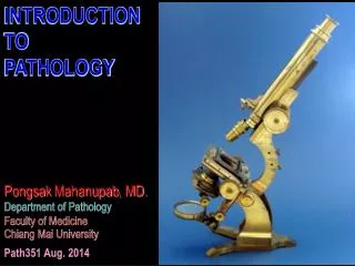
Department of Pathology Faculty of veterinary medicine. Viral diseases BY Dr. SHEREIN SAEID. VIRAL DISEASES. Introduction.

E N D
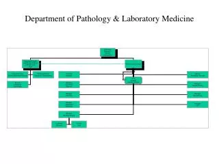
Department of Pathology Faculty of veterinary medicine
* Viruses can be classified into two classes DNAand RNA viruses.* Most viruses stimulatelymphocytes, however, equine encephalomyelitis stimulateneutrophilsfirstly andthen lymphocytes.* Most viruses result incoagulative necrosis, however, equine encephalomyelitis result inliquifactive necrosis.
Virus and tissue reaction
* Some viruses resulting in lysis and necrosis of cells---- -----------------------------Herpes v.*Some viruses resulting in Proliferation of cells---------- -----------------------------Pox,Leukosis.*Some viruses resulting in Fusion of cells and syncytial formation-------------Paramyxovirus.*Some viruses resulting in Apoptosis of cells-------------- ----------------------------Rift valley fever.*Some viruses resulting in Transformation of cells--------- --------------------------Lung adenomatosis.*Some viruses resulting in Inclusion bodies which either – ----------------intranuclear ( I/N )-----------Herpes. ------------------intracytoplasmic ( I/C )------Pox. ------------------I/N and I/C---------------Paramyxovirus.
Virus and infection
* Acute infection: Virus is eliminated by host immune response. * Chronic or persist infection: Virus is not eliminated and can be isolated for long time.* Latent infection: Virus is apparently eliminated and reappears under stress factors as herpes.
Immunotolerant animal:*Animal get infected by virus and considered it as self antigen(no antibodies)*Animal appeared normal but sheds virus to other animalsAs in BVD infection.
Viral diseases characterized by vesicle formation
Foot and mouth diseaseF.M.DAphthus feverAphthusruminitisDEF.* Viral disease of cloven footed animals; cattle, sheep, camel, and may pig.* Not affect horse.* Ch’ch’ : Vesicle formation on the buccal cavity, interdigital space, rumen, reticulum, omasum, and udder.CausePicornaviridae(epitheliotropic)R.N.AR.O.IIngestion – InhalationPathogenesisVirus ( via R.O.I )………………………………….. Buccal cavity ( str. Spinosum ) …………………………………………Ballooning degeneration(hydropic)…………………………Vesicle( aphthus )or bullae ……………………………………………….Erosion ……………………….. Ulcer…………………………………………..Virus to circulation ………………………………………………………………..Viremia …………………………….G.I.T, Interdigital space & udder.
Lesions (i) MACRO* Buccal lesions : -Vesicle or bullae on anterior 2/3 of the dorsum of tongue. -Dental pad and buccal cavity are also affected.* Foot lesions: -Affect interdigital space ………………………………..Lamness.-In complicated cases the claws may slaughed.* G.I.T lesions: -Vesicles, Necrosis & Erosions in oesophagus, rumenAPHTHUS RUMINITIS., reticulum, omasum,and intestine. -Peticheal H. on abomasums, intestine, and subendocardium.* Udder lesions : Vesicle on Teat extend to Mamaryts …………………………………….Mastitis.(ii) MICRO*Epidermis -Ballooning (hydropic Degenerations) -Vesicle(serous fluid,epithelialcells,inflame.cells(mostly lymphocytes & fewneutrophil………………………………………..Erosion …………………………………………………Ulcer * Dermis Dermatitis i) Congestion ii) Inflammatory cells ( lymphocytes + neutrophils) iii) Perivascular cuffing
Malignant F.M.DDEF.: *Per acute F.M.D of young calves end with rapid death due to(ACUTE GENERAL VENOUS CONGESTION ).*Ch’ch’ non suppurativemyocarditis. Cause :R.N.A virus.R.O.I :Ingestion & Inhalation. Pathogenesis :Virus via R.O.I……Viremia ….Heart muscles & Skeletal muscles.Lesions :(i) MACRO :Greyish necrotic foci on the wall of left ventricle take TIGER appearance SO known as TIGER HEART.(The same lesions are seen in skeletal Ms.)ii)MICRO :Zenker’s necrosis + Inflammatory cells ( Lymphocytes + Macrophages ).(The same lesions are seen in skeletal Ms.)***F.M.D In SHEEP :Vesicle on teat, vulva, & dental pad////// Milder than in cattle//////// F.M.D In Lambs & Pigllets Usually DIE.
Vesicular stomatitis Vesicular exanthema • DEF.:* • Viral disease affect mainly horses. • * May affect cattle and pig. • * Ch’ch’ vesicle on the buccal cavity, • Interdigital space and udder • * Seasonal disease as it occurs in • summer coz of mosquito. • * Enzootic disease as it occurs in • U.S.A • Cause: • Rhabdoviridae genus (aphthous) e’ 7 Ag types(A,O,C,sat1,sat2,sat3,AsIA1) • e’out cross protection between them • R.O.I • Mosquito bite • wound contamination • Def.: • * Viral disease affect • mainly pigs. • * Ch’ch’ vesicle on the • buccal cavity, • Interdigital space • And udder • Cause: • Calciviridae • R.O.I: • Ingestion
Vesicular stomatitis Vesicular exanthema • Pathogenesis • Virus ( via R.O.I ) ………Buccal cavity ( str. Spinosum)……….. spongiosis………………. Vesicle ( aphthus ) or bullae… Erosion…………Ulcer. • Virus to circulation ……… Viremia …… ……………………………G.I.T, Interdigital space & udder. • Lesions: • As F.M.D ( Buccal, Foot, and Udder Lesions ONLY) • N.B • (HYDROPIC DEGENERATION REPLACED BY SPONGIOSIS IN HORSE VESICULAR STOMATITIS)
DIAGNOSIS OF FMD,Vesicularstomatitis&Vesicular exanthema:(1) Clinical Signs (Fever(aphthus fever), Off food, Salivation,Smaking of mouth,vesicles on its sites.(2) Post mortem lesions(Macro and Micro).(3) Isolation And IdenificationOf Virus.(4) Diffrential diagnosis from other vesicular diseases.Control measures :(1)Complete eradication of infected animal. (2) Attenuated vaccine for only 1 virus to avoid cross immunity.
PoxViridae virus
GENERAL Characters : • (i) Type : D.N.A virus with 6 genera; Orthopox, Capripox, Lapripox, Suipox, Avipox, and Parapox. • (ii) Tropism : Epitheliotropic. • (iii) R.O.I : Insect bite, Wound contamination (contact ), Inhalation, Ingestion, and • Coitus. • (iv) Animal susceptiple : • * Affect most animals except dogs. • * Horse pox----------- 3 forms; Leg, Buccal, and Genital . Ch’Ch’ Pock lesions . • * Cow & Buffalo …….mild affection. …. Affect teat & udder causing pock lesions. • Microscopic lesions as those of sheep. • * Camel …………Fatal in Young ………… Local in adult. Ch’Ch’ Pock lesions . • * Human … • (i) Pseudo cow pox ( MILKER’S NODULES ) • Transmitted to milkers from teat cow pox through milking. • (ii) Small pox ( VARIOLA ) • Fatal as it has systemic reaction + skin affections. • Human vaccinized against small pox.
(v) Lesions : Cutanous & Systemic lesions. • (vi) Inclusions : I/C inclusion bodies ( GUARNIERI BODIES ) • (viii) Pathogenesis of sheep pox lesions In order : • (i) ROSEOLA St. …….. Red line of dilated vessel. • (ii) MACULA St. …….Edema & Inflammatoty exudates. • (iii) PAPULE St. ………Proliferations of cells. • (iv) VESICULAR St. ……Vesicle formation. • (v) Pastule St. • (vi) UMBLICATED St. ( POCK LESION )…………….. • vesicles ruptured giving ulcer with elevated borders. • (vii) ENCRUSTATION St……..Crust with scab formation. • (viii) HEALING St. …………..Healing of deep pitted ulcer.
Sheep pox - under tail (elevated papule) previous quit
Sheep pox (scab formation) previous quit