Download
1 / 26
260 likes | 601 Views
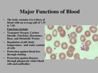
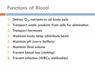
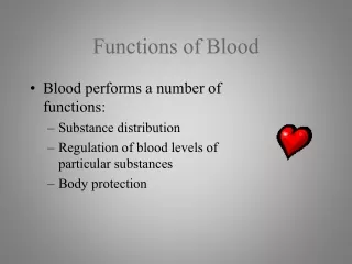
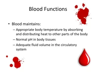
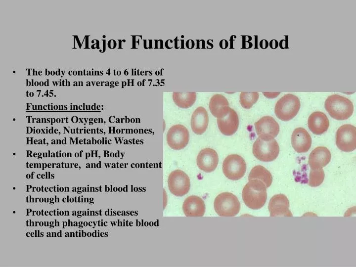
Major Functions of Blood. The body contains 4 to 6 liters of blood with an average pH of 7.35 to 7.45. Functions include : Transport Oxygen, Carbon Dioxide, Nutrients, Hormones, Heat, and Metabolic Wastes Regulation of pH, Body temperature, and water content of cells

E N D
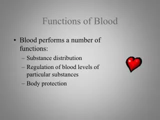
Major Functions of Blood • The body contains 4 to 6 liters of blood with an average pH of 7.35 to 7.45. Functions include: • Transport Oxygen, Carbon Dioxide, Nutrients, Hormones, Heat, and Metabolic Wastes • Regulation of pH, Body temperature, and water content of cells • Protection against blood loss through clotting • Protection against diseases through phagocytic white blood cells and antibodies
Components of Normal Adult Blood Measured via Hematocrit: female 38% to 46%, male 40% to 54%
Abnormal Hematocrits • Polycythemia: Increased RBC production. • Physiologic Polycythemia: Increase in RBC production due to hypoxic tissues, like what occurs at high altitudes. • Polycythemia Vera: genetic mutation in the hemocytoblastic cell line that increases RBC production. Hematocrit values can reach 70%
Formation of Blood cells - Hemopoiesis Hormones: RBC: Erythropoietin Platelets: Thrombopoietin WBC’s: Colony-stimulating Factors (4)
Red Blood Cells or Erythrocytes • Contain hemoglobin to carry oxygen • 4.8 to 5.4 million RBC’s per microliter • Produced at a rate of 2 million RBC’s per second • Lack a nucleus • Biconcave to increase surface/volume ratio • Diameter of 7 – 8 microns
RBC Shape and the Structure of Hemoglobin • RBC’s pass through capillary beds in single file. • Hemoglobin is made of four polypeptides: Two Alpha and two Beta that contain a heme unit • Each heme can carry an O2 • There are about 280 million hemoglobin molecules in each RBC • Other molecules such as CO2 and NO are also carried by hemoglobin
Changes in hemoglobin • Anemia: A deficiency of RBCs, which can be caused by either too rapid loss or slow production. • Blood loss Anemia: Due to hemorrhage, plasma is replaced in 1-3 days, but, RBC replacement takes longer. • Microcytic Hypochromic Anemia: Low levels of hemoglobin in RBCs due to chronic blood loss resulting in low Fe3+ levels in newly produced RBCs. • Aplastic Anemia: Decreased RBC production in bone marrow due to chemical, drug, or radiation exposure. • Pernicious Anemia: Chronic illness caused by impaired absorption of Vitamin B-12 because of a lack of intrinsic factor (IF) in gastric secretions. Vitamin B12, in turn, is necessary for the formation of red blood cells.
Changes in Hemoglobin • Anemia: A deficiency of RBCs, which can caused by either too rapid loss or slow production. • Hemolytic Anemia: Different abnormalities of RBCs that make RBCs fragile and rupture easily. • Hereditary Spherocytosis: RBC develop as small spherical cells rather than being biconcave. These spherical cells easily rupture by slight compression. • Sickle-cell Anemia: Genetic mutation causing abnormal beta chains. When this hemoglobin is exposed to low O2 concentrations, it precipitates into long crystals that cause the cells to become sickle-shaped.
Hemolytic Disease of Newborns (HDN)or Erythroblastosis Fetalis
WBC Anatomy and FunctionNeutrophil • Make up 60 to 70% of WBC’s • 10 –12 um. In diameter • Nucleus 2-5 lobes (increase with cell age) • Fine granular cytoplasm • Phagocytic cells the engulf bacteria • Increase: stress, burns, bacterial infections • Decrease: Radiation exposure, B12 deficiency
WBC Anatomy and FunctionEosinophil • Make up 2-4% of WBC’s • 10 –12 um in diameter • Nucleus 2–3 lobed • Cytoplasm filled with large red granules • Combat histamines in allergic reactions • Phagocytic on antigen/antibody complexes • Destroy certain parasitic worms • Increase: allergic reactions, parasitic infections, autoimmune disease
WBC Anatomy and FunctionBasophil • Make up .5 to 1% of WBC’s • 8- 10 um in diameter • Cytoplasm filled with large deep blue-purple granules • Liberate heparin and histamines during allergic reactions • Intensify inflammatory response • Increase: Allergic reactions, leukemia, cancers, hypothyroidism • Decrease: Pregnancy, ovulation, stress, hyperthyroidism
WBC Anatomy and FunctionMonocytes • Make up 3-8 % of WBC’s • 12 – 20 um in diameter • Nucleus is kidney-shaped • Cytoplasm is non-granular • Phagocytic cells • Increase: Viral and fungal infections, tuberculosis, and some leukemias • Decrease: Bone marrow depression, treatment with cortisol
WBC Anatomy and FunctionLymphocytes • Make up 20 to 25% WBC’s • Small: 6-9 um in diameter Large: 10-14 um • Nucleus is round or slightly indented • Cytoplasm forms rim around nucleus • B cells produce antibodies • T cells attack viruses, cancer cells, and transplanted tissues • Natural killer cells attack infectious microbes and tumor cells
WBC Anatomy and FunctionLymphocytes • Increase: Viral infections and some leukemias • Decrease: Prolonged illness, immunosuppression
Platelet Anatomy and Function • Disc-shaped 2 – 4 um in diameter • 150,000 to 400,000 per uL of blood • Alpha Granules: contain clotting factors, platelet-derived growth factor • Dense Granules: contain ADP, ATP, Ca2+, serotonin, and fibrin-stabilizing factor
Steps in Vascular Damage and Clotting Responses • Vascular Spasm: contraction of smooth muscle in arteriole walls to reduce blood flow. • Platelet plug formation: 1. Platelet adhesion: Platelets contact and stick to free collagen fibers of the damaged blood vessel 2. Platelet release reaction: Activated platelets extend many projections that enable them to contact and interact with one another. They then liberate their granules.
Steps in Vascular Damage and Clotting Responses Liberated ADP and thromboxane A2 help activate other platelets. Serotonin and thromboxane A2 function as vasoconstrictors helping to decrease blood flow. 3. Platelet aggregation: Liberated ADP makes new platelets sticky; these newly-recruited and activated platelets adhere to the originally-activated platelets. The process causes the formation of a platelet plug.
The Blood Clotting Cascade • Extrinsic Pathway (Fast acting) 1. Tissue Factor (TF) or Thromboplastin is released by tissue cells outside of the damaged vessel. 2. TF begins a chemical reaction pathway that activates Thrombokinase (F10). F10 combines with Proaccelerin (F5) to form the enzyme Prothrombinase.
The Blood Clotting Cascade • Intrinsic Pathway (slow acting) Activated by factors within the blood or vessels Antihemophilic factor D or Hageman factor (F12) is activated by contact with collagen fibers. F12 starts a chemical cascade that ultimately activates F10 or Thrombokinase. F10 combines with Proaccelerin (F5) to form the enzyme Prothrombinase.
The Blood Clotting Cascade • The Common Pathway Prothrombinase catalyzes the conversion of Prothrombin (F2) to Thrombin. Thrombin converts the soluble plasma protein fibrinogen in the insoluble protein fibrin (loose threads). Thrombin also activates Fibrin Stabilizing Factor (F13) which converts the loose threads into stable threads.
The Problems with Clotting Cascade • Hemophilia A: Deficiency of Factor VIII accounts for 85% cases. • Almost exclusively in males. Females are usually carriers • caused by a gene mutation on the “X” chromosome. Occurs in about 1/10,000 male births • Other Hemophilias account for another 15% • Hemophilia B (Factor IX) • Hemophilia C (Factor XI) • Hemophilia D (Factor XII)
The Problems with Platelets and Abnormal Clotting • Thrombocytopenia: Abnormally low levels of platelets. Usually below 50,000/ul of blood. • In many cases, specific antibodies are produced against platelets destroying them • Thrombus: Abnormal clot that develops in a blood vessel. • Embolus: Free thrombic clots carried in the blood that usually get caught in arterioles in the brain, kidney, and lungs.