Download
1 / 68
780 likes | 1.04k Views
Common Running Injuries. Robert P. Wilder, MD, FACSM Chair, Physical Medicine & Rehabilitation The University of Virginia Medical Director, The Runner’s Clinic at UVA Team Physician, Ragged Mountain Racing. Objectives. Identify common contributors to running injuries

E N D
Common Running Injuries Robert P. Wilder, MD, FACSMChair, Physical Medicine & RehabilitationThe University of VirginiaMedical Director, The Runner’s Clinic at UVATeam Physician, Ragged Mountain Racing
Objectives • Identify common contributors to running injuries • Describe treatment for heel pain, stress fractures, and patellofemoral pain syndrome • Understand the importance of proper mechanics in managing injury • Outline criteria for running while treating injury
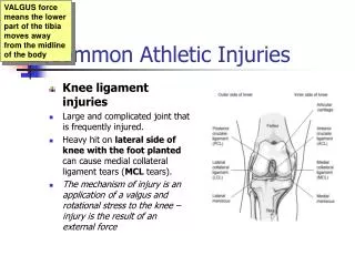
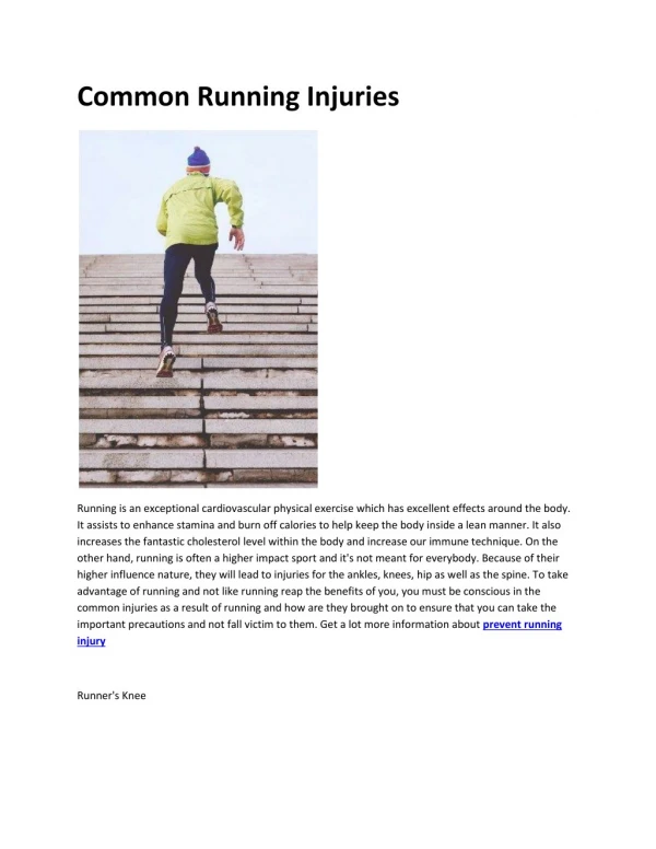
Epidemiology of Running Injuries 30 million active runners 70% all runners sustain significant injury 40% knee 15% each: shin, achilles, hip/groin 10% foot and ankle 5% spine 25% recreational 5% elite
Epidemiology of Running Injuries 4% bit by dogs 0.3% hit by bicycles 0.6% hit by cars 7% hit by thrown objects
Intrinsic Abnormalities Malalignment Muscle imbalance Inflexibility Muscle weakness Instability
Extrinsic Abnormalities Training errors Equipment Environment Technique Sport-imposed deficiencies
Examination of the Injured Runner History Biomechanical assessment Site-specific exam Dynamic exam Shoe exam Ancillary testing radiologic electrodiagnostic compartment testing
History • Prior injury history • Team/Club • Identify transitions • MPW (20, 40) • Long run (< 1/3 weekly total) • Intensity • Surface (? Muscle tuning) • Shoes/orthotics (350-400 miles) • Cross Training • Goals • Life Stressors/fatigue • Females: eat d/o, menstrual irreg, osteopenia
Physical Examination • Biomechanical assessment • Site specific examination • Dynamic examination • Ancillary testing • Shoe examination
Functional Screening • Single Leg Stance • Single Leg Squat • Bilateral Squat • FHB isolation • Step-down Test • STAR Excursion Test • Swing Test
Functional Screening Single Leg Stance
Functional Screening Single Leg Squat
Functional Screening Bilateral Leg Squat
Functional Screening FHB Isolation
Functional Screening Step-Down Test
Functional Screening STAR Excursion Test
Functional Screening Swing Test
Plantar Fasciitis • 10% U.S. Population • 600,000 outpatient visits annually • 7-9% all running injuries
Plantar Fascia • Thick aponeurosis • Arises from medial calcaneal tuberosity • Spans arch • Bands circle flexor tendons • Insert proximal phalanx
Functions During Gait Cycle • Heel strike: Allows midfoot to become flexible, absorb shock, conform to uneven surface • Toe off: Windlass Mechanism: Shortening increases arch, locks midtarsal, stabilizes toe off
Pathophysiology • Overuse • Inflammation • Chronic changes (collagen necrosis, angiofibroplastic hyperplasia, chondroid metaplasia, matrix calcification) • Tearing • Medial vulnerable (thin, limited vascular supply, limited ability to stretch
Risk Factors • Obesity • Excessive time on feet • Limited ankle motion (tibiotalar) • Limited great toe mobility (extension) • Inflexibility (HS and achilles) • Pes cavus • Pes planus • Leg length inequality (short leg)
Presentation • Plantar heel pain • A.M. pain • Mid arch (sprinters) • Increased pain with running • Imaging primarily to rule out other causes
Treatment • Relative Activity Modification • Anti-inflammatories • Flexibility (HS, gastroc-soleus, plantar fascia) • Manual therapy (ankle and great toe mobility: tibiotalar subtalar, great toe) • Strength (Foot intrinsics, ankle stability, lower quarter stability)
Treatment (cont) • Devices – CTF brace, heel cushions • Low dye taping • Night splints and socks • Inserts • Steroid injections
Treatment (cont) • ESWT (> 12 mos) • Botulinum A • Autologous blood • PRP • Prolotherapy
Recalcitrant Cases • Confirm diagnosis • Surgical release • 75-95% “some improvement” • 27% significant pain • 20% activity restriction • Fasciectomy + neurolysis of nerve to ADM • Percutaneous plantar fasciotomy • Flouroscopically-assisted fasciotomy • US guided fasciotomy
Heel Pain Differential • Fat Pad Insufficiency • Calcaneal Stress Fracture
Heel Pain Differential (cont) • Neuropathies • Tarsal Tunnel Syndrome • Medial plantar nerve (“Joggers Foot”) • First Branch, Lateral Plantar nerve (“Baxter’s Neuropathy”) • Radiculopathy
Heel Pain Differential (cont) • Tendonopathies • PTTD (posterior tibial) • Flexor • Peroneal • Achilles
Heel Pain Differential (cont) • Spring Ligament injury
Heel Pain Differential (cont) • Bursitis • Pre-achilles • Retrocalcaneal
Heel Pain Differential (cont) • OS Trigonum Syndrome (differentiate from posterior talus fracture)
Heel Pain Differential (cont) • Haglund’s
Heel Pain Differential (cont) • Sever’s Syndrome (kids)
Heel Pain Differential (cont) • Achilles enthesopathy (consider inflammatory)
Heel Pain Differential (cont) • Tarsal coalition
Heel Pain Considerations • Ankle mobility (tibiotalar, subtalar great toe) • Flexibility (HS, GS, PF) • Ankle stability • Lower quarter stability
Stress Fractures Failure of bone to adapt adequately to mechanical loads (ground reaction forces and muscle contraction) experienced during physical activity • Tibia • Metatarsals • Fibula • Navicular
Stress Fractures (cont) • Non-critical (relative rest 6-8 wks) • Medial tibia • Metatarsals 2,3,4
Stress Fractures (cont) At risk fractures: • Femoral neck • Anterior tibia • Medial malleolus • Navicular • Base 5th metatarsal
Femoral Neck Superior (distraction) – higher incidence worsening/ non union Inferior – (compression)
Anterior Tibia Casting vs relative rest up to 6-8 months If no healing – ortho (transverse drilling, grafting, medullary fixation)
Navicular • Tender N-spot • Critical zone middle 1/3 • Non-weight bearing 6-8 weeks • Progressive activity over 6 more weeks
Proximal 5th Metatarsal • Jones fx of proximal diaphysis • Cast 6-10 weeks • Non-union: ortho • Consider ortho early in competitive • Contrast with avulsion: symptomatic RX