Download
1 / 16
160 likes | 166 Views
Aim : How can we identify and describe the human body cavities?. DO NOW:. A. Body Cavities. Two essential functions: Protect organs from accidental shocks Permit changes in size and shape of internal organs Ventral body ( Front/anterior cavity) Divided by the diaphragm: Thoracic cavity

E N D
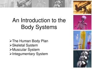
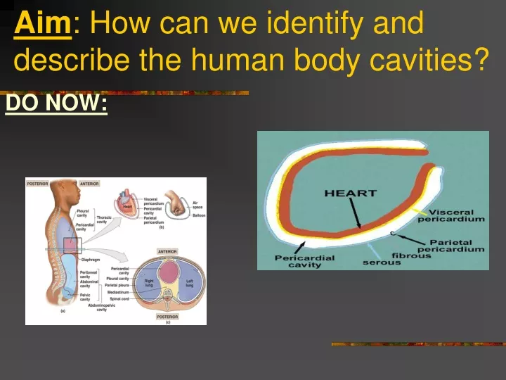
Aim: How can we identify and describe the human body cavities? DO NOW:
A. Body Cavities Two essential functions: Protect organs from accidental shocks Permit changes in size and shape of internal organs Ventral body ( Front/anterior cavity) Divided by the diaphragm: Thoracic cavity Abdominopelvic cavity
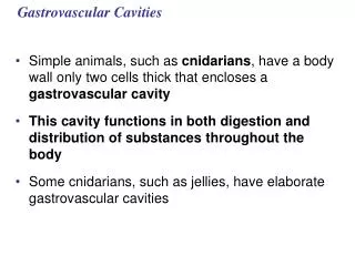
B. Viscera Organs of the thoracic and abdominal pelvic cavities Serous membrane is a thin slippery membrane that covers the viscera Parts of the serous membrane: Visceral layer Covers the viscera within the cavities Parietal layer Lines the wall of the cavities
C. Serous membrane of the thoracic cavity Pleura-Refers to the lungs Serous membrane of the pleural cavities Visceral pleura clings to surface of lungs Parietal pleura lines the chest wall Pericardium- Refers to the heart Serous membrane of the pericardial cavity Visceral pericardium covers the heart Parietal pericardium lines the wall of the pericardial cavity.
D. The Thoracic Cavity Separated into regions Right and left pleural cavities contain right and left lungs Mediastinum upper portion filled with blood vessels, trachea, esophagus, and thymus lower portion contains pericardial cavity the heart is located within the pericardial cavity
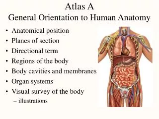
Body Cavities FIGURE 1–11 The Ventral Body Cavity and Its Subdivisions.
E The Abdominopelvic Cavity 1. Peritoneal cavity — chamber within abdominopelvic cavity Parietal peritoneumlines the internal body wall Visceral peritoneumcovers the organs
Abdominal cavity — superior portion Diaphragm to top of pelvic bones Contains digestive organs Retroperitoneal space Area posterior to peritoneum and anterior to muscular body wall Contains pancreas, kidneys, ureters, and parts of the digestive tract
Pelvic cavity — inferior portion Within pelvic bones Contains reproductive organs, rectum, and bladder
Body Cavities FIGURE 1–10 Relationships Among the Subdivisions of the Ventral Body Cavity.
F. Dorsal Body Cavity- Posterior Cranial cavity Protects the brain Vertebral canal Formed by bones of vertebral column Contains the spinal cord Meninges Layers of protective tissue that line the cranial cavity and the vertebral canal.
Other Cavities Oral (mouth) cavity Tongue and teeth Nasal cavity nose Orbital cavities eyeball Middle ear cavities Small bones of the middle ear Synovial cavities Joints
SUMMARY Create a Concept Map that illustrates today’s topic.