Download
1 / 19
190 likes | 211 Views
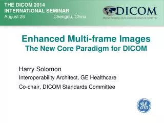
DICOM in Ophthalmology, an Example of a New Enhanced Multiframe Object. Herman Oosterwijk. Add logo if desired. Enhanced Multiframe. Objective: This presentation will answer the following question:

E N D
DICOM in Ophthalmology, an Example of a New Enhanced Multiframe Object Herman Oosterwijk Add logo if desired
Enhanced Multiframe • Objective: This presentation will answer the following question: • What are the major features of the new generation enhanced multiframe DICOM objects, how to understand this new type of object, and how does a typical enhanced multiframe look like, in particular OCT (Optical Coherence Tomography)
Enhanced multiframe: • Agenda: • Major features of the new generation of DICOM objects, i.e. enhanced multiframe • Steps to encode/decode this new type of object • OCT object, a sample enhanced multiframe
Traditional Multiframe objects: Vector • Single image, multiple frames • US, NM, XA, RF, SC, VL • Simple structure: • # frames • Frame rate • Frame index pointer • Frame increment pointer (NM)
Enhanced multiframe: • Why a new generation of objects: CT, MR, ophthalmology, mammography tomo, XA, RF • Imaging is moving from pixels in slices (2-D) to voxels (3-D) and beyond (n-D), i.e. time, space, frequency, flow: functional imaging (CTA, etc.) • Simple “Study-Series-Image” hierarchy does not suffice anymore for organization, viewing, browsing, sorting using the appropriate hanging protocols • Text descriptions for Series leaves room for guessing
Enhanced multiframe: • Solution: enhanced multiframe • Many Attributes are required (Type 1) • Where possible, Attribute contents are encoded • Definition of multiple dimensions • Header size reduction • Protocol efficiency: 1000 slice CT • 1000 times C-Store RQ…RS vs 1 time RQ…RS • ONE structure serving as a template for all new SOP Classes (CT, MR, XA, RF, OCT, MG…)
5 5 5 4 4 4 3 3 3 2 2 2 1 1 1 Time (1) Space (2) Dimension example: 3 \ 1 \ 3 Dimension Index Values 5 Trigger Delay Time ID 1 Stack ID=1 100 ms • Dimension • Indexes (3): • Trigger Delay Time • Stack ID • Position 50 ms ID 2 1 0 ms ID 3 Time
Enhanced multiframe encoding: • Fixed per frame: • Name, ID…. • Accession #, Study ID… • KV, mAs…. • Orientation…. • #rows, columns, bits • Split header: • Fixed information • Variable information (keep as minimum as possible) • Some information can be in both places (contrast) Header Part 1 • Variable for one/more frames: • Position • Time • Scab type Header Part 2 Pixels in frames
Enhanced multiframe: • Header part 2 (variable) definition: • Implemented as “functional groups”: SQ with zero, one, or more items • Documented as “macros” in Multi-Frame Functional Group Module • Common functional groups, used for multiple modalities, such as “Frame Content Macro” • Structure of dimensions, and concatenations is “generic”: Multi-frame functional group and dimension modules • Modality specific functional groups, e.g. for MR, ophthalmology, etc.
Multiframe Functional Groups: Shared attributes Per-frame attributes Pixel data
Ophthalmology: IOD:
OPHTHALMIC TOMOGRAPHY FUNCTIONAL GROUP MACROS
Enhanced multiframe: • Implementation issues: • Support by vendors, especially PACS archives and even more workstations: growing, but slowly • Mixed environments: new and old, especially for established modalities (CT, MR, XA, RF) • Handling very large files: concatenation (not allowed for OCT) – splitting up into mini-multi frames • Retrieval on frame level: to be added to standard
Conclusion: • New, enhanced multiframe objects are defined in DICOM, some for existing, some for new modalities • These new objects provide better interoperability for n-D (esp. dynamic) objects, allow for more sophisticated display protocols, and give better performance • Modalities, and to a lesser degree PACS systems are starting to support it; a mixed environment is inevitable
Thank you! Herman Oosterwijk: herman@otechimg.com www.otechimg.com