Download
1 / 35
350 likes | 354 Views
In these lectures, we explore the use of biochemistry and genetics to define the function of DNA polymerase, focusing on fidelity and specificity. We break down complex processes into intermediates and subreactions, analyze the players and activities involved, and detect and identify intermediates. By structurally characterizing intermediates, we can identify the proteins and nucleic acids responsible for the activities. We also examine the replication fork and replisome, and explore the intricacies of DNA replication.

E N D
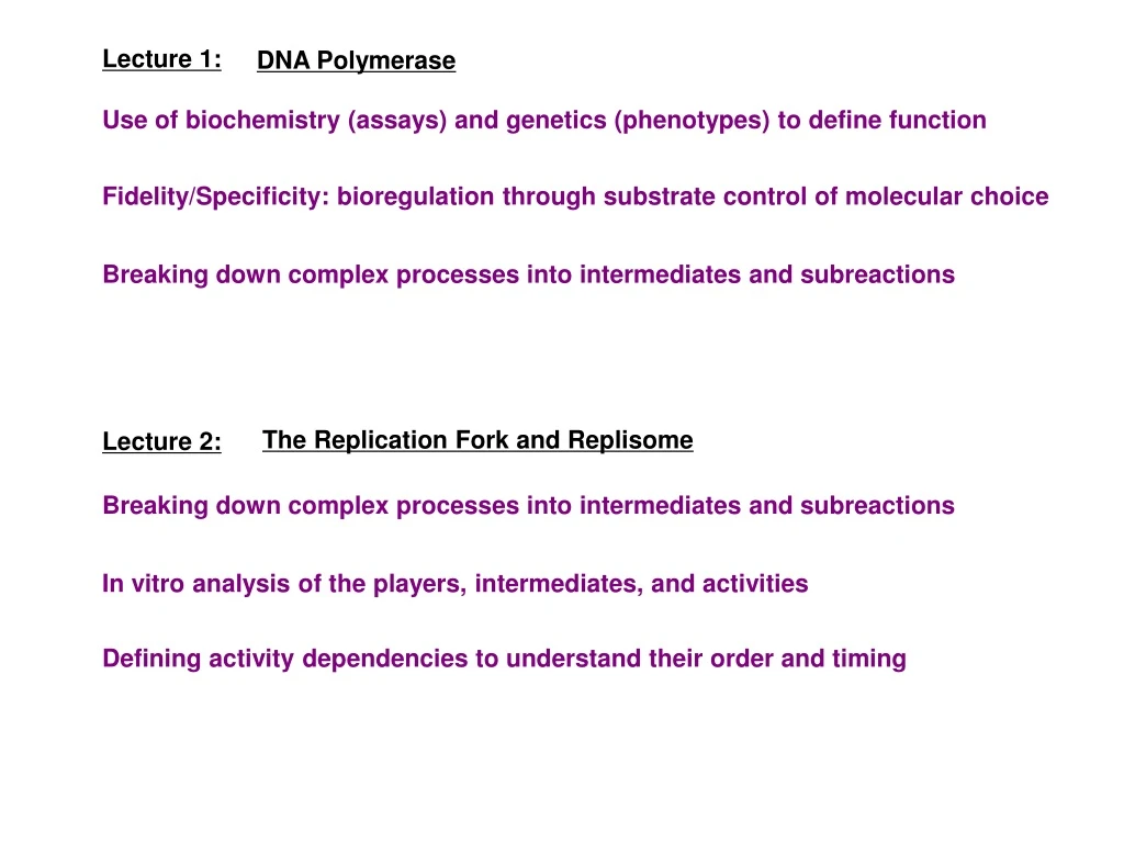
Lecture 1: DNA Polymerase Use of biochemistry (assays) and genetics (phenotypes) to define function Fidelity/Specificity: bioregulation through substrate control of molecular choice Breaking down complex processes into intermediates and subreactions The Replication Fork and Replisome Lecture 2: Breaking down complex processes into intermediates and subreactions In vitro analysis of the players, intermediates, and activities Defining activity dependencies to understand their order and timing
A1 A2 A3 A4 An+1 S I4………….. In P I1 I2 I3 5’ 3’ 3’ 5’ 5’ 3’ Dissecting Complex Molecular Mechanisms S = substrate S P P = product I = intermediate A = activity
A1 A2 A3 A4 An+1 S I4………….. In P I1 I2 I3 Dissecting Complex Molecular Mechanisms S = substrate S P P = product I = intermediate A = activity How to detect and identify intermediates? How to structurally characterize intermediates? How to identify the proteins/nucleic acids responsible for the activities?
HL HL HH Visualization of E. coli DNA Replication Intermediates daughter Label E. coli ~ 2 generations with radioactive thymidine (H ) 3 fork Gently lyse cells and let DNA settle and stick onto a membrane parent fork Autoradiograph with coating of photographic emulsion daughter Develop emulsion and analyze DNA structures under microscope, quantifying lengths E. coli genome is circular and replicates with a replication bubble containing two equally long daughter arms connected at each end to the remaining parental segment Infer double-strand labeling (HH) vs single-strand labeling (HL) from quantification of silver grain density DNA replication is localized to two moving replication forks that travel bidirectionally around the molecule probably from a single site of initiation
Dissecting Complex Molecular Mechanisms How to detect and identify intermediates? Detecting highly abundant intermediates by precursor labeling How to structurally characterize intermediates? Direct visualization of single molecules by microscopy How to identify the proteins/nucleic acids responsible for the activities?
Reconciling polymerase directionality with antiparallel DNA strands Onestrand: 5’>3’ polymerase can move continuously in same direction as replication fork Other strand: 5’>3’ polymerase must move discontinuously in opposite direction as replication fork 5’ Fork Movement 3’ 3’ 5’ 5’ 3’ Is there a transient intermediate where newly synthesized DNA is in “short” single strands? Is one of the daughter molecules single-stranded near the fork ?
time time P S I1 I2 I1 P S I2 I1 I1 I2 S I2 P S I2 I2 I1 I1 P P P S P I1 I2 I2 Examples of blocks: Single molecule analyses use similar strategy but - remove/inactivate protein - remove cofactor • do not require synchronization • - do establish molecular fate - lower temperature - add inhibitor Detecting Intermediates Synchronize Reaction To Transiently Enrich Successive Intermediates Pulse-Chase Label a Synchronous Cohort Block Reaction Step To Accumulate Intermediate S P S I1 I2 I1 S { Partial Reaction Molecular fate suggested by block and established if reversing block converts I to P Molecular fate suggested by temporal transitions Molecular fate established by chase Label can enhance sensitivity and specificity of detection
A1 A2 A3 A4 An+1 S I4………….. In P I1 I2 I3 Dissecting Complex Molecular Mechanisms S = substrate S P P = product I = intermediate A = activity How to detect and identify intermediates? How to structurally characterize intermediates? How to identify the proteins/nucleic acids responsible for the activities?
Structural Analysis of Intermediates Examples of structural features that can be monitored Nucleic Acids Complexes Proteins Composition Cofactor (NTP) Status Size Conformation Stoichiometry Shape Modifications Conformation DS versus SS Interacting Sequences Ligand Binding Strand Pairing Interacting Domains Covalent Linkages Strand Polarity Covalent Linkages Modifications Topology Sequence
Detection and Analysis of Newly Synthesized DNA Label replicating E. coli for seconds with H -thymidine 3 Extract DNA and alkali denature Centrifuge in alkaline sucrose gradient to separate by size Measure radioactivity in gradient fractions (increasing size ) The newest DNA synthesized is mostly small (~ 1000-2000 bp) In another paper, 10-20% of the label chased into large DNA EM visualization of fork by Inman showed SS DNA on one arm Structural analysis by others showed 8-10 nt RNA at 5’ end
Semi-Discontinuous DNA Synthesis Leading strand: polymerase moves continuously in same direction as replication fork Lagging strand: polymerase moves discontinuously in opposite direction as replication fork 5’ D 3’ C Fork Movement Lagging B 3’ 5’ A Leading 5’ 3’ Additional activities inferred from replication intermediate analysis A. helix unwinding B. priming Okazaki fragment synthesis & processing C. primer replacement prokaryotes: 1–2 kb eukaryotes: 100–200 bp D. ligation
Using in vitro (soluble cell-free) Systems • The advantages of an in vitro system for understanding mechanism • How one validates an in vitro system • How one can purify the activities in the in vitro system • How one can use the purified system to understand its activities
A1 A2 A3 A4 An+1 S I4………….. In P I1 I2 I3 Advantages of an in vitro system to study mechanism Can isolate a process from other competing or disruptive processes S = substrate S P P = product I = intermediate A = activity How to detect and identify intermediates? Easier to synchronize, pulse-label, or block the process How to structurally characterize intermediates? Easier to isolate and structurally analyze intermediates Easier to introduce various defined intermediates (or substrates) How to identify the proteins/nucleic acids responsible for the activities? Can separate and purify activities without any a priori knowledge about them
Example: replication elongation DS DNA template; dNTP replication fork okazaki fragment replication mutants aphidicolin (for eukaryotes) fork rate okazaki fragment size Validating an in vitro system Show the in vitro system shares many properties of the in vivo process Substrate Product Intermediates Genetic Requirements Inhibitor Sensitivity Quantitative Properties
Purifying biochemical activities from in vitro systems In Vitro Complementation Fractionation & Reconstitution Can accelerate by trying to replace fractions with suspected proteins purified from expression systems
Phage T4 DNA Replication in vitro in vivo in vitro 800 nt/sec 500 nt/sec Fork Rate ~ 2 kb ~ 2 kb Okazaki Fragment No OF maturation Genetic Requirements 32, 41, 43, 44, 45, 62 32, 41, 43, 44, 45, 62 Biochemical activities mostly purified by in vitro complementation Can reconstitute reaction with seven purified activities
A direct assay for helicase activity * * 41 is NOT required for rapid synthesis on SS DNA 41 has GTP/ATPase activity Greatly stimulated by SS DNA Inhibition by GTPS slows strand displacement synthesis A Helix Unwinding (Helicase) Activity 41 is required for rapid strand displacement synthesis on DS DNA SLOW no 41 FAST no 41 FAST
Replicative Helicases Form hexameric rings that encircle single-stranded DNA and hydrolyze ATP to translocate unidirectionally along the DNA Prokaryotes 5’ > 3’ (on lagging strand): DnaB Discussion Paper Eukaryotes 3’ > 5’ (on leading strand): Cdc45-Mcm2-7-GINS 5’ 5’ 5’ 3’ 3’ 3’ 5’ 5’ 5’ DnaB 3’ 3’ 3’ Belong to AAA+ ATPases family, which form multimeric complexes and couple ATP binding and/or hydrolysis to conformational changes
Activities for okazaki fragment maturation (E. coli) DNA Pol I (5’>3’ exo) Excise Primer DNA Pol I Fill-In Gap Ligase Seal Nick
Replication Fork Tasks and Activities Leading Strand Task Activity polymerase synthesize DNA helicase separate parental strands primase prime polymerase nuclease/polymerase replace primer ligase connect okazaki fragments SSBP stabilize SS DNA clamp loader/clamp ensure processivity Lagging Strand unlink parental strands topoisomerase
A1 A2 A3 A4 An+1 S I4………….. In P I1 I2 I3 Understanding Molecular Mechanisms Some activities may affect the rate, fidelity, specificity, or regulation of these steps S = substrate S P P = product I = intermediate A = activity How to detect and identify intermediates? How to structurally characterize intermediates? How to identify the proteins/nucleic acids responsible for the activities?
Processivity How many times an enzyme can act repeatedly on a substrate before dissociating from it Assay: measure product size under conditions where an enzyme cannot reassociate with its substrate once it dissociates Condition 1: preload enzymes onto substrates then dilute Condition 2: excess substrate (e.g. primer-template) distributive polymerase (not processive) processive polymerase
An activity that enhances polymerase processivity 44/62 ATPase and 45 enhance the processivity of T4 DNA polymerase 43 Continuous ATP hydrolysis by 44/62 is not required for enhanced processivity Once ATP is hydrolyzed, processivity factors act like a “sliding clamp” for the polymerase
A1 A2 A3 A4 An+1 S I4………….. In P I1 I2 I3 Understanding Molecular Mechanisms S = substrate S P P = product I = intermediate A = activity How to detect and identify intermediates? How to structurally characterize intermediates? How to identify the proteins/nucleic acids responsible for the activities? How is proper order and timing of activities maintained?
The Challenge of Regulating and Coordinating Multiple Activities Primase synthesizes primer Clamp-loader positions clamp around primer-template Polymerase loads onto primer-template and binds to clamp Primase synthesizes primer for next okazaki fragment Polymerase synthesizes okazaki fragment What regulates where and when primers are made? Polymerase dissociates from clamp to load onto next primer Clamp-loader loads clamp What regulates polymerase processivity? Okazaki fragment maturation is completed Clamp-loader eventually releases clamp for reuse on other okazaki fragments What directs when clamps are released? Adapted from Molecular Biology of the Cell. 4th Ed.
Keeping the Lagging Strand Polymerase at the Replication Fork Processive synthesis of okazaki fragments by lagging strand polymerase suggests tethering to leading strand replication proteins at the fork, generating a dynamic lagging strand loop (trombone model). In E. coli, tau dimer tethers by binding two core polymerases in the Pol III holoenzyme B clamp core • Complex clamp-loader t dimer core B clamp Pol III holoenzyme Predicted lagging strand “loop” seen in EM; dynamic loop behavior detected by single molecule analysis Figures from Molecular Biology of the Cell. 4th Ed.
Trombone Model from Cell Snapshots (Cell 141:1088) How many polymerases can interact with each clamp? How do primase and helicase interact yet work in opposite directions? What holds leading and lagging strand polymerases together in other systems? Are leading and lagging polymerization coordinated? See Movie at http://www.youtube.com/watch?v=4jtmOZaIvS0
Replication forks must deal with many problems and dangers Many genomic insults are now thought to originate from replication accidents DNA lesions induce responses to: (1) protect stalled forks (2) bypass lesions (3) delay further initiation (4) block cell cycle 2 3 1 4 Segurado & Tercero, Biol. Cell (2009) 11:617-627
Appendix Bioreg 2015 Replication Lecture 2
HL HL HH Full interpretation of the Cairns theta structure daughter D fork parent fork daughter At the time label was added the great grandparent molecule, which had initiated from an origin near the bottom left corner, had replicated all but the region from C to D (marked by arrowheads). As this round of replication was completed the resulting grandparent molecule became labeled on one strand just between C and D Initiation and completion of the next round of replication generated the parent molecule with one strand fully labeled and the other (inherited from the grandparent molecule) labeled only from C to D. Thus, the molecule is labeled on both strands between C and D and This parent molecule was then caught in the act of replicating bwith two thirds of it replicated by forks X and Y, generating two daughter arms labeled A and B. Arm A was derived from the mostly unlabeled parental strand and is thus mostly labeled only on the new daughter strand (except from D to X) . Arm B was derived from the labeled parental strand and is thus labeled on both strands.
Modifying Okazaki’s Fully Discontinuous Synthesis Model Okazaki: newly synthesized DNA is mostly small suggesting discontinuous replication on both strands Smith & Whitehouse (2012): inactivate ligase in Saccharomyces cerevisiae sequence small SS DNA see opposite strand bias on either side of origins Inman & Schnos (1971): electron microscopy of replicating phage l DNA SS is often seen on only one arm of each fork In some cases interrupted by short DS segment DS SS DS SS DS DS SS DS SS DS Thus, there is in vivo evidence supporting semi-discontinuous DNA synthesis (see slide notes)
Summary of Activities and Proteins at the Replication Fork Note: Many of these activities are also required for DNA repair or recombination, and in several cases the same proteins are used Diagram shows prokaryotic 5’>3’ helicase on lagging strand 3’>5’ eukaryotic helicase would be placed on leading strand E. coli Eukaryotes Activity Task DnaB Mcm2-7, Cdc45, GINS unwind parental strands helicase primase DNA Pol a-primase prime DNA synthesis primase SSBP RPA1-3 stabilize SS DNA SSBP * ** DNA Pol III core DNA Pol e, DNA Pol d synthesize DNA polymerase ensure processivity clamp loader, clamp g-complex, b subunit RFC1-5, PCNA ? coord leading and lagging t subunit Ctf4? unlink parental strands topoisomerase Topo I/Gyrase, Topo IV Topo I/Topo II replace primer polymerase/nuclease DNA Pol I/RNaseH DNA Pol d, FenI, Dna2 connect okazaki fragments ligase DNA Ligase DNA Ligase I * DNA Pol III Holoenzyme ** leading, lagging
E. Coli Clamp-Loader (gdd’) loads the Clamp (b ) onto DNA through the ordered execution of activities, each of which is dependent on the intermediate generated by the previous activity 3 2 Key Interactions Order Activities Clamp Loading Model d alone can bind and open clamp interface d’ binds d and blocks interaction with clamp (sequesters d in the clamp-loader) g has ATPase activity ATP binding induces conformational change in g and releases d from d’ (allows g to bind and open clamp) Clamp binding inhibits g ATPase (prevents premature clamp release) Clamp binding enhances clamp-loader binding to primer-template ( promotes clamp delivery to DNA) • Primer-template binding stimulates • g ATPase (allows g to release and • close clamp to complete loading) Energetics Clamp opening depends on protein-ATP (g - ATP) and protein-protein (b - d) binding energies Clamp closing depends on ATP hydrolysis