Download

1 / 31
420 likes | 1.16k Views
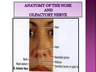
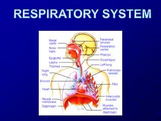
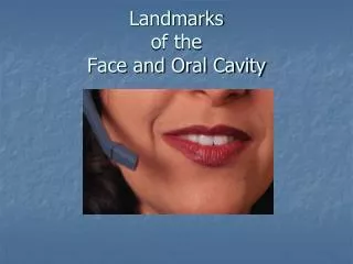
Nose, Nasal cavity & Paranasal Sinuses. Dr. Zeenat Zaidi. Nose. root. Only externally visible part of the respiratory system Has a free tip and is attached to the forehead by the root or the bridge

E N D
Nose, Nasal cavity & Paranasal Sinuses Dr. ZeenatZaidi
Nose root • Only externally visible part of the respiratory system • Has a free tip and is attached to the forehead by the rootor the bridge • Has two openings, the anterior (external) nares or nostrils, which lead to the nasal cavity • Each nostril is bounded laterally by the ala and medially by the nasal septum tip ala septum external nares
Nose: Structure • Nose consists of bony & cartilaginous framework • Formed above by the: • Nasal bones • Frontal processes of maxillae • Nasal part of frontal bone • Formed below by plates of hyaline cartilage, which include upper & lower nasal cartilages and the septal cartilage Nasal part of Frontal bone
Nasal Cavity • Extends from the external (anterior) naresto the posterior nares (choanae) • Divided into right & left halves by the nasal septum • Each half has a: • Floor • Roof • Lateral wall • Medial wall (septum)
Roof Is narrow & formed (from behind forward) by the: Body of sphenoid Cribriform plate of ethmoid bone Frontal bone Nasal bone & cartilage Floor Separates it from the oral cavity Formed by the hard (bony) palate
Medial Wall (Nasal Septum) • Osteocartilaginous partition, only rarely lying in the midline • Covered by the mucoperiosteum • Formed: • Superiorly by the vertical (perpendicular) plate of ethmoid bone • Posteriorly by the vomerbone • Anteriorly by the septal cartilage
Lateral Wall • Shows three horizontal bony projections, covered by mucous membrane, the superior,middle&inferiorconchae (turbinates) • The superior and middle conchae are parts of the ethmoid bone, whereas the inferior concha is a separate bone • The cavity below each concha is called a meatus and are named as superior, middle & inferior corresponding to the conchae
The small space above the superior concha is called the sphenoethmoidal recess The middle meatus is continuous in front with a depression called theatrium Atrium is limited bove by a ridge called agar nasi Below and in front of atrium, and just within the nostril lies thevestibule
The conchae increase the surface area of the nasal cavity The recess & meati receive the openings of the: Paranasal sinuses Nasolacrimal duct
Sphenoethmoidal recess: Receives the opening of the sphenoidal sinus • Superior meatus:Receives the opening of the posterior ethmoidalsinus • Inferior meatus:Receives the opening of thenasolacrimal duct. The opening is guarded by a valve, a fold of mucous membrane
Middle meatus: • Shows a rounded eminence, the ethmoidal bulla, caused by the bulging of the underlying middle ethmoidal sinus, which opens on its upper border. • A curved groove, hiatus semilunaris, lies below the bulla. Hiatus receives the opening of the maxillary sinus • Anterior end of hiatus leads to funnel-shapedinfundibulum, which receives the openings of the frontal & the anterior ethmoidal sinuses
Lining of the Nasal Cavity • Vestibule is lined by modified skin, and has short, curved hair called vibrissae • The roof, upper part of the septum, upper surface of the superior concha, and the sphenoethmoidal recess are lined by the olfactory mucosa • The rest of the cavity is lined by the respiratory mucosa A V V
Olfactory Mucosa • Contains olfactory cells (bipolar sensory ganglion cells), which serve as receptors for olfactory stimuli. • Distinct smells are far more numerous than tastes • The sense of smell plays a major role in the flavor of foods and it is common for individuals who lose their sense of smell to report that food loses its taste. (food seems somewhat tasteless when a person has cold) • Most air breathed in normally flows through the nose but only a small part reaches the olfactory mucosa, enough to get a response to an odor. Sniffing, however, increases the flow of air over the smell receptor cells, greatly increasing their exposure to odors.
Respiratory Mucosa • Pseudostratified ciliated columnar epithelium with goblet cells • Rests on thick network of thin walled veins that warms the air as it flows through the cavity • Glands produce ‘mucus’, which: • moisten the air • cleans the air by trapping the incoming bacteria and foreign debris • Cilia help in moving the contaminated mucus posteriorly towards the throat, where it is swallowed and digested by the stomach juices
Nerve Supply • Nasal cavity receives sensory& visceralinnervation Sensory innervation • Olfactory mucosasupplied by olfactory nerves • Nerves of general sensation are derived from opthalmic & maxillary nerves • Anterior part supplied by the anterior ethmoidal nerve (branch of opthalmic nerve) • Posterior part supplied by nasal, nasopalatine and palatine branches (of maxillary nerve)
Visceral Innervation Sympathetic fibers arise from neurons of superior cervical ganglionand are distributed through plexuses around the arteries, supply mainly vascular smooth muscle Parasympathetic fibers arise from neurons of the pterygopalatine ganglion that course in the nasopalatine nerve (branch of maxillary) and its branches, supply the mucosal glands.
Arterial Supply • Sphenopalatine artery (branch of the maxillary artery) is the main supply • Alar and septal branches of superior labial artery (branch of the facial artery) • Anterior & posterior ethmoidal arteries (branches of the ophthalmic artery) • The arteries make a rich anastomosis in the region of the vestibule, and anterior portion of the septum
Venous Drainage: Veins begin as a rich plexus in the submucosa, accompany the corresponding arteries, and drain into the facial, ophthalmic, and sphenopalatine veins. LymphaticDrainage: The lymphatics from the: Vestibule drain into the submandibular lymph nodes Rest of the cavity drains into the upper deep cervical lymph nodes
Functions of Nose & Nasal Cavities • Air conditioning: warming, cleaning and humidifying the inhaled air • Add resonance to the voice • Vocal sounds are also produced in the nasal cavity thus aiding in vocalisation • Involved in the special sense of smell • Central role of the nose in facial appearance • ??
Paranasal Sinuses • Air filled cavities located in the bones around the nasal cavity: ethmoid, sphenoid, frontal bones & maxillae • Lined by respiratory mucosa which is continuous with the mucosa of the nasal cavity • Drain into the nasal cavity through relatively small apertures • Drainage of the sinuses mainly depends on the movement of the cilia, which propel the mucus toward their openings in the nasal cavity
The development of sinuses begins in 3-4 month, but only maxillary & ethmoidsinuses are present in rudimentary form at birth. The frontal & sphenoidal sinuses are not clinically perceptible at birth and can rarely be demonstrated on plain x-ray before two years of age. Continue to grow postnatally Enlarge appreciably after 8th year & become fully formed at adolescence E M From a 3 months old fetus, showing ethmoid & maxillary sinuses
Functions • Lighten the skull • Act as resonant chambers for speech • The respiratory mucosal lining helps in warming, cleaning and moistening the incoming air
Maxillary Sinuses • Located within the body of the maxilla • Pyramidal in shape with the base forming the lateral wall of nose & the apex lies in the zygomatic process of the maxilla • Roof: formed by the floor of the orbit • Floor: formed by the alveolar border. Roots of 1st and 2nd premolars and the 3rd molar (sometimes canines) project into the sinus • Opens into the middle meatus through the hiatus semilunaris • Supplied by superior alveolar & infraorbital nerves M
Frontal Sinuses • Two in number • Located within the frontal bone, separated from each other by a bony septum • Triangular in shape, extending backward into the roof of the orbit • Opens into the middle meatusthrough the infundibulum • Supplied by the supraorbital nerve
Ethmoidal Sinuses • Locatedwithin the ethmoid bone, between the nose and the orbit • Divided into three groups: anterior, middle & posterior • Anterior group opens into the infundibulum, middle opens on the bulla, and posterior into the superior meatus • Supplied by the anterior and posterior ethmoidal nerves
Sphenoidal Sinuses • Two in number • Located within the body of sphenoid • Open into the sphenoethmoidal recess • Supplied by the posterior ethmoidal nerve
Clinical Notes • Epistaxis:Little’s area, common site of bleeding from nose • Inflammation of the nasal mucosa, Rhinitis, results in nasal congestion and excessive production of mucus leading to ‘postnasal drip’ • Infections of the nasal cavity can extend to the: • Paranasal sinuses • Nasolacrimal duct & lacrimal sac
Inflammation of mucosa of the sinuses, Sinusitis, causes excessive production of mucus leading to obstruction of the drainage of sinuses. This results in headache and change in the voice Infection of frontal & anterior ethmoidal sinus can easily spread to maxillary sinus because of the location of their openings Infection of upper teeth can lead to inflammation of the maxillary sinus Extraction of an infected upper tooth may result in a fistula
The maxillary sinus is most commonly the site of infection The inflamed mucosa results in excessive production of mucus as well as narrowing of its opening in the nasal cavity The position of the drain causes problems in that mucus can collect in the sinus below the drain. In this situation, the sinus will only drain if the patient lies on their opposite side. Pressure from the trapped fluid/mucus causes sinus pain