Download
1 / 1
10 likes | 85 Views
Trace Elemental Composition and Concentration of Upstate New York Rainwater Samples Using the Union College Pelletron Particle Accelerator and Proton Induced X-ray Emission Spectroscopy Katie Schuff , Maria Battaglia , Scott LaBrake , Colin Gleason , Charles Harrington, & Michael Vineyard

E N D
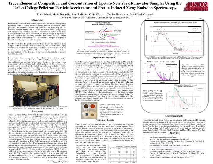
Trace Elemental Composition and Concentration of Upstate New York Rainwater Samples Using the Union College Pelletron Particle Accelerator and Proton Induced X-ray Emission Spectroscopy Katie Schuff, Maria Battaglia, Scott LaBrake, Colin Gleason, Charles Harrington, & Michael Vineyard Department of Physics & Astronomy, Union College, Schenectady, NY Introduction Environmental pollutants from various sources, both natural and anthropogenic, have been found to deposit harmful elements into our environment. These pollutants are generally airborne and eventually find their way into the watershed and soils through rainfall. Plants and animals uptake these pollutants and in high enough quantities are toxic. Environmental pollutants are known to have harmful effects on the human body [1]. Thus it is of great importance to be able to detect and accurately determine the amount of these harmful pollutants and in addition understand the deposition, transport and uptake of these elements by organisms in the ecosystem. In order to identify the specific elements found in aerosol, rainwater or soil samples, and thus determine their concentrations, the non-destructive , highly sensitive, and accurate ion beam analysis technique of Proton Induced X-ray Emission Spectroscopy (PIXE) will be used. PIXEallows for a fast and accurate method for the detection of environmental pollutants in aerosols, rainwater, and soils. In particular, rainwater samples will be collected from various geographic locations throughout upstate New York and Union College’s particle accelerator was used to generate a 2.0-MeV proton beam. Spectra of x-ray intensity versus x-ray energy will be collected and a PIXE analysis will be performed. The presence and concentration of selected trace elemental pollutants will be determined and preliminary results will be presented. Future studies will be performed to further explore the possible sources of these pollutants. In particular an elemental map showing the distribution of elements as a function of geographic area will be created. Figure 2: Drawing showing the basics of the PIXE process [3]. Here the ground and first excited states of an arbitrary atom are shown. The inner shell electron is ejected by the proton and a higher orbital electron transitions from the n = 2 to the n = 1 state with an emission of an x-ray photon. This is called a Ka transition. Figure 4: PIXE Spectra of dried Rainwater deposited on Mylar Film for three different geographic regions in upstate New York and overlaid with the PIXE spectrum for the Mylar foil alone. Experimental Procedure Rainwater samples were collected in June, July, and September 2009 from the Historic Stockade District located in Schenectady, NY, Clifton Park, NY and Hadley, NY. Following the method of Ghorai et. al., [5] approximately 200-mL of rainwater were obtained and dried in order to prepare a target that can be put under vacuum. The rainwater solutions (where only the Schenectady, NY sample consisted of an internal standard of 1-mL of selenium added to the 200-mL rainwater sample producing a concentration of 5,000-ppm) were slowly evaporated, in glass that had been acid washed, at approximately 70-80°C until 1-mL of the sample remained. Selenium was chosen as an internal standard as selenium is known not to be an element found within rainwater [5]. This remaining 1-mL was deposited onto an approximately 12-mm thin Mylar film, shown in the bottom of Figure 3, and further dried in a vacuum desiccator creating a thin film target that was run in the particle accelerator. X-rays produced by the incident proton beam were collected by a silicon drift detector and the resulting spectra of intensity versus x-ray energy were analyzed using GUPIX[4], thus allowing for the determination of the trace elemental composition and concentration of the sample. In order to calibrate GUPIX [4]a set of Micromatter standards[6] were used as shown in the top of Figure 3. The concentrations of these standards was used to normalize the concentrations of the elements found in the rainwater. Figure 5: On the left are shown PIXE spectra for Mylar foil with deposited rainwater (blue) and blank Mylar foil (pink). The two overlaid spectra provide a clear visualization of the trace elemental composition of the rainwater without any effects due to the Mylar foil. Figure 6: On the right are PIXE spectra for the Schenectady NY rainwater deposited on Mylar Film (green dots) and the fit generated by GUPIX[4] (red curve) allowing for the determination of the elemental composition and concentration. Figure 3: Photograph of two of the Micromatter standards [6], in the upper left and right, used for calibration of GUPIX[4]. In addition the photograph shows the non-rainwater deposited Mylar film on the bottom left and a rainwater deposited Mylar film on the bottom right. Figure 1: Photograph of the Union College tandem electrostatic Pelletron particle accelerator used to generate the 2.0-MeV proton beam that will be used to facilitate the PIXE process. Experiment Union College‘s tandem electrostatic Pelletron particle accelerator, shown in Figure 1, was used to generate a 2.0-MeV beam of protons that was steered to be incident onto a dried rainwater target and Q = 10-mC of charge was collected. When the protons interact with elements in the target there is a probability, called the cross-section, that inner shell electrons will be ejected and that higher orbital electrons will de-excite to fill these vacancies accompanied by an emission of an x-ray photon. This is called Proton Induced X-ray Emission Spectroscopy (PIXE) [2]and a schematic is shown in Figure 2 [3]. Each element has its own unique set of x-ray energies associated with electron transitions and this allows us to fingerprint each element. To fingerprint the elements we generally look for K-series x-rays, which are electron transitions from any orbital higher than the ground state to the ground state of the atom. The lower energy, higher probability n = 2 to n = 1 transition is termed a Ka transition, while the higher energy, lower probability n = 3 to n = 1 transition, is called a Kb. X-ray spectra are collected on a silicon drift detector and are displayed as intensity versus x-ray energy and are analyzed to determine the trace elemental composition of the target with a software program called GUPIX[4]. Using trace elemental standards, GUPIX [4]will be calibrated to give concentrations of selected trace elements in the rainwater target. GUPIX [4] calculates the concentrations Ctarget from the x-ray yields of the sample and target, Ysample and Ytarget, the amount of charge collected, Q, the x-ray detector efficiency, e and an overall energy independent constant, H shown in the equation below. Acknowledgements I would like to thank Union College and in particular the Department of Physics and Astronomy for presenting me with this opportunity to further explore physics and in helping me find my own path in the field. Also, I would like to thank NASA’s NY Space Grant for their financial support and of course my research group. I would like to thank my advisor Prof. Scott LaBrake for all of his time and patience and also Maria Battaglia, Colin Gleason, Chad Harrington and Prof. Mike Vineyard for their time and effort with this project. Preliminary Results Figure 4 shows the raw data collected by the x-ray detector for 3 different geographic regions in upstate New York and a non-rainwater deposited Mylar foil are displayed as the intensity of the x-rays as a function of their energy. Figure 5 shows the raw data for the Schenectady, NY rainwater sample and Mylar film overlaid against the non-rainwater deposited Mylar film for comparison. Examination of figure 6, which shows the GUPIX fit to the Schenectady NY rainwater spectrum allows one to identify at least 10 trace elements and using GUPIX [4]their respective concentrations are calculated and are given in the table below. These concentrations have been normalized using the Micromatterstandards[6] . References The Environmental Protection Agency http://www.epa.gov/climatechange/effects/health.html Particle Induced X-ray Emission Spectroscopy. Johannson, S. Campbell, J. Malmgvisit, K. Wiley, NY 1995 The University at Albany, State University of New York, http://ibl.albany.edu/research/techniques/pixe.html GUPIX, University of Guelph. http://pixe.physics.uoguelph.ca/gupix/main/ Trace Elemental Composition and Acidity of Central-Alabama Rain. Ghorai, S.K. Tekyi-Mensah, O. Sims, J.F. Williams, J.R. Alford, W.L. NIM B, 63, 139-142. MicromatterCo. 18218 18th Ave NW Arlington, WA 98223 By comparing the three spectra we can visibly see the trace elemental composition of the rainwater without any effects of the Mylar foil. Determining the trace elemental composition allows us to further analyze and determine the concentrations of these various elements using GUPIX [4] as a function of geographic area.