Download
1 / 12
120 likes | 369 Views
Improving Corneal Symmetry & Visual Acuity in Keratoconus with Keratometry-Guided Conductive Keratoplasty Prior to Intrastromal Corneal Ring Implantation. Combined CK and Intacs for Keratoconus: Improved Corneal Symmetry and Visual Acuity. Anita Nevyas-Wallace, MD Bala Cynwyd, Pennsylvania.

E N D
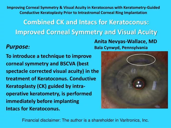
Improving Corneal Symmetry & Visual Acuity in Keratoconus with Keratometry-Guided Conductive Keratoplasty Prior to Intrastromal Corneal Ring Implantation Combined CK and Intacs for Keratoconus:Improved Corneal Symmetry and Visual Acuity Anita Nevyas-Wallace, MD Bala Cynwyd, Pennsylvania Financial disclaimer: The author is a shareholder in Varitronics, Inc. Purpose: To introduce a technique to improve corneal symmetry and BSCVA (best spectacle corrected visual acuity) in the treatment of Keratoconus. Conductive Keratoplasty (CK) guided by intra-operative keratometry, is performed immediately before implanting Intacs for Keratoconus.
Method Report of 3 cases of keratoconus treated with 4 - 6 CK spots placed at the 9mm optical zoneimmediately beforeimplantation of a singleIntacssegment (Addition Technology, Des Plaines, IL) in the maximally elevated hemi-meridian. Pressing with a blunt instrument mimics the effect of corneal steepening and shrinkage from a CK spot. CK spot placement is determined by pressing on the corneain different hemi-meridians and assessing which most circularizes the egg-shaped reflection of the “ring of lights” of the operative keratometer (Varitronics, Inc., Broomall, PA). Successive spots are placed (maximum six), until slight overcorrection is observed.
Ways CK Has Been Used in Keratoconus • Personal communication • Personal communication • Previously, CK has been used to steepen the flat meridian (Boxer Wachler1) [place CK spots in flat area] More recently, • To shrink and “centralize” the cone (Hardten2) [place CK spots in most elevated area] • The method described here shrinks and centralizes the cone under keratometric guidance
Customizing CK Application Pattern Varitronics Operative Keratometer with Movable Fixation Device • Elevation Topography • Crucial for identifying most elevated hemi-meridian • Operative Keratometry • Intraoperative feedback
Intraoperative Feedback is Key in CK for Keratoconus • Instrument pressure mimics the corneal steepening and shrinkage of CK • This guides placement of each CK spot Ring reflection assessed as blunt instrument presses different areas
Technique For CK Before Intacs Identify “point” of egg-shaped reflection Determine where pressure with blunt instrument best circularizes the egg shape, then place CK spot Repeat steps 3 and 4 Pachymetry: Verify > 500 μ at 9 mm o.z. of most elevated hemi-meridian Mark 9 mm optical zone (o.z.)
Technique For CK Before Intacs • Once point of egg is slightly flattened with CK (4 to 6 spots), then • Make tunnels & Insert Intacs
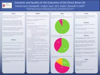
Elevation difference map Pre-Intacs with CK Post-op Post Cone much flatter, less peripheral after CK with Intacs Results All patients showed improvement in UCVA and BSCVA of at least 2 lines (Snellen). Corneal symmetry on elevation corneal topography was improved in all patients.
Cone much flatter, less peripheral after CK/Intacs Pre-Op Elevation maps Curvature maps Post-Op GJ – o.d.
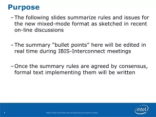
Cone becomes flatter, less elevated, more central Cone much flatter, less peripheral after CK/Intacs Elevation maps Curvature maps 3 month Pre-op 6 month CP
Conclusion Guided by operative keratometry and performed just before implantation of Intacs for keratoconus, Conductive Keratoplasty flattens, shrinks and displaces centrally the cone so that the Intacs segment may be placed in a more effective position for improving corneal symmetry and for reducing regular and irregular astigmatism in keratoconus.