Download
1 / 6
80 likes | 444 Views
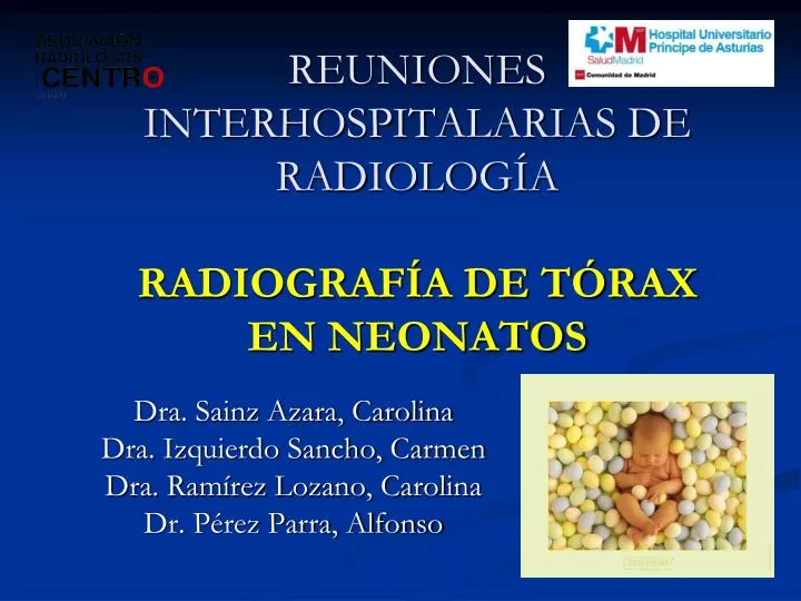
REUNIONES INTERHOSPITALARIAS DE RADIOLOGÍA RADIOGRAFÍA DE TÓRAX EN NEONATOS. Dra. Sainz Azara, Carolina Dra. Izquierdo Sancho, Carmen Dra. Ramírez Lozano, Carolina Dr. Pérez Parra, Alfonso. Información clínica. RNPT de 29 + 1 semanas. Antecedente materno: Preeclampsia . Parto: cesárea.

E N D
REUNIONES INTERHOSPITALARIAS DE RADIOLOGÍARADIOGRAFÍA DE TÓRAX EN NEONATOS Dra. Sainz Azara, Carolina Dra. Izquierdo Sancho, Carmen Dra. Ramírez Lozano, Carolina Dr. Pérez Parra, Alfonso
Información clínica • RNPT de 29 + 1 semanas. • Antecedente materno: Preeclampsia. • Parto: cesárea. • PAEG (940gr). • Leve distrés respiratorio a su llegada a neonatos. • Empeoramiento clínico a la hora y media de su llegada con aumento del trabajo respiratorio y necesidad de FiO2. • Analítica: acidosis mixta.
¿Cómo definiría el patrón radiológico? • Patrón reticulonodular. • Broncograma aéreo. • Derrame pleural bilateral. • Bullas enfisematosas. • 1 y 2 son correctas. ¿Cuál de las siguientes NO es correcta? • Se trata de una enfermedad típica de los RN a término. • Se debe a un déficit de surfactante pulmonar. • Una radiografía normal a las 6 horas del nacimiento prácticamente descarta el diagnóstico. • Una de sus complicaciones es el enfisema intersticial pulmonar.
¿Cuál de las siguientes NO es correcta? • Resolución de periférico a central. • Resolución de LLSS a LLII. • Pueden existir atelectasias. • Puede existir hemorragia pulmonar. • Es característico el derrame pleural. ¿Cuál es el diagnóstico? • Neumonía bacteriana. • Enfisema intersticial pulmonar. • Enfermedad de la membrana hialina. • Secuestro pulmonar.
ENFERMEDAD DE LA MEMBRANA HIALINA • También llamado síndrome de distrés respiratorio pulmonar (SDR). • RN < 36 semanas y < 2,5 kg. • Síntomas comienzan poco después del nacimiento. • Retracción anormal del tórax, cianosis y aumento FR. • Se debe a déficit de surfactante pulmonar. • Función: Disminuye la tensión superficial dentro de los alveolos evitando la atelectasia. • Rx: Patrón reticulonodular. Broncograma aéreo, atelectasias e hipoventilación. • Evolución: Continua ingresada. • Bibliografía: Kirks. Radiología pediátrica.