Download

1 / 34
340 likes | 408 Views
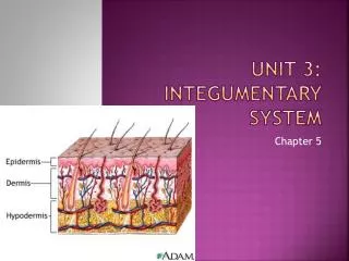
The Integumentary System Unit-D. Explain the structures of the Integumentary System Layers of Skin. 1H04.01. EPIDERMIS Outermost layer of the skin. 2 (of 3) epidermal layers are: · Stratum corneum · Stratum germinativum. Outermost layer of the epidermis

E N D
The Integumentary System Unit-D
Explain the structures of the Integumentary System Layers of Skin 1H04.01
EPIDERMISOutermost layer of the skin • 2 (of 3) epidermal layers are: • ·Stratum corneum • ·Stratum germinativum
Outermost layer of the epidermis In cells, cytoplasm replaced by KERATIN – making them waterproof. ¨Flat and scale-like cells that flake off. ¨First line of defense against surface bacteria. Repair itself if injured Thickest on palms of hands, soles of feet STRATUM CORNEUM
Innermost epidermal layer ¨Reproductive layer – cells form and push their way up, become keratinized, and replace the top layer Contains MELANOCYTES – cells that contain a pigment = MELANIN STRATUM GERMINATIVUM
1. Black, brown, or has a yellow tint – depending on racial origin 2. The more melanin, the darker the skin 3. Caucasians don’t have much melanin in their melanocytes. 4. Freckles = patches of melanin 5. Albinism = no melanin Exposed to sunlight causes a darken of melanocytes. MELANIN
Ridges in stratum germinativum that arise from dermis Create permanent ridges in fingers, palms and soles of feet These “friction ridges” help with grip & Cause “fingerprints” PAPILLAE
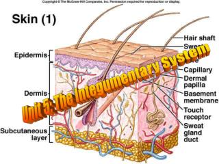
Thicker inner layer that contains: 1. Connective tissue 2. Blood vessels 3. Nerve endings 4. Muscles 5. Hair follicles 6. Oil and sweat glands 7. Fat cells DERMIS
·Sensory nerves – heat, cold, touch, pain and pressure ·Touch receptors close to the surface ·Pressure receptors are deeper NERVE RECEPTORS IN DERMIS
·Lies under the dermis (not really part of integumentary system) ·Made up of loose connective tissue Contains half of the body’s stored fat so undergo the most change with wt. gain. SUBCUTANEOUS LAYER
HAIR ·Almost everywhere on the body ·Length, thickness, type and color varies Outer layer = CORTEX Inner layer = MEDULLA Part under the skin = ROOT Visible part of hair is the = SHAFT FOLLICLE = hollow tube in the dermis, which hair grows in. PAPILLA = tuft of tissue in root, contains capillaries APPENDAGES OF THE SKIN
ARRECTOR PILI MUSCLE = smooth muscle attached to follicle. Causes the skin to pucker when exposed to a chill. NAILS ·Nail is formed in the nail bed or MATRIX Epidermal cells fused together and fill with keratin APPENDAGES OF THE SKIN
·Perspiration is 99% water ··Distributed over the entire skin surface ·Large numbers AXILLA under the arms, palms of hands, soles of feet and forehead Under arm odor caused by bacteria mixed with sweat. ·Duct extends to form a pore in the skin, perspiration excreted through the pores ·May be activated by heat, pain, fever and nervousness ·Average fluid loss is 500 ml per day SWEAT GLANDSSUDORIFEROUS GLANDS
SEBACEOUS GLANDS • Oil (SEBUM) producing gland of the skin that protects and lubricates.
1H04.02 • Analyze the functions of the Integumentary System
Skin = Integument = Cutaneous Membrane 7 Functions: 1.Protective covering 2.Regulates body temperature 3.Manufactures Vitamin D 4.Sensory function 5.Temporary storage of fat, glucose, water and salts 6.Screens out harmful ultraviolet radiation 7. Absorbs certain drugs Topical drugs-lotions, birth control patches ect.. INTEGUMENTARY SYSTEM
Intact skin = best protection from germ invasion, pathogens, toxins and water loss Skin generally too dry for microbial growth – they do grow in moist areas Most skin bacteria associated with hair follicles or sweat glands Best way to prevent spread of infection of disease is handwashing PROTECTION
1H04.03 • Discuss characteristics & treatment of common skin disorders.
DISORDERS OF THE SKIN • ACNE • ¨Common and chronic disorder of sebaceous glands • ¨Sebum (oil) plugs pores, deposits hardens & pores are plugged, fills with leukocytes. • ¨Also – blackheads, cysts, pimples and scarring
Normal hair is replaced by a very short, transparent hair. Alopecia/Baldness
DISORDERS OF THE SKIN • ATHLETE’S FOOT • ¨Contagious fungal infection • ¨Usually contracted in public baths and showers • ¨Rx – anti-fungal agents
DISORDERS OF THE SKIN • DERMATITIS • ¨Non-specific inflammation of skin • ¨Can be rash – reaction to soap, plants, substance. • ¨Can be emotional – stress can cause skin blotches
DISORDERS OF THE SKIN • GENITAL HERPES • ¨Viral • ¨Blister in genital area • ¨Spread through sexual contact • ¨Periods of remission and exacerbation • ¨Rx – Acyclovir • ¨Can be passed to newborn during vaginal delivery
TANNING • Sunlight stimulates melanocytes to make more melanin • Tanning produced by UV rays. • Primary cause of skin cancer is direct sunlight.
SKIN CANCER ¨Associated with exposure to sun (UV rays) • Most common type of cancer in people
DISORDERS OF THE SKIN • MALIGNANT MELANOMA • ¨Occurs in melanocytes • ¨Metastasizes to other areas quickly • ¨Appears as brown or black irregular patch that occurs suddenly • ¨A change in an existing wart or mole may indicate melanoma • ¨Rx – surgical removal of melanoma and surrounding area and chemotherapy
1. Benign: Not dangerous. 2. Biopsy: Removal of a small piece of tissue for examination under a microscope. 3. Lesion: A change in the structure or appearance of a part of the body as the result of an injury or infection. 4. Malignant: Threatening to life. Skin Cancer: Words
FIRST DEGREE BURN • ¨Superficial • ¨Skin red and dry • ¨Involves only epidermis • ¨Rx – cold water • Healing within one week
SECOND DEGREE BURN • ¨Epidermis and dermis • ¨Pain, swelling, redness and blistering • Subject to infection • ¨Skin may be exposed to infection • ¨Rx – pain medication, dry sterile dressing • Healing within 2 weeks
THIRD DEGREE BURN • Epidermis, dermis and subcutaneous layers (full thickness) • Loss of skin, blackened skin • May be life threatening • Rx-prevention of infection, fluid replacement, skin grafting
RULE OF NINES – Measures percent of body burned. Body divided into 11 areas, each is 9% of body surface.