Download
1 / 54
630 likes | 1.45k Views
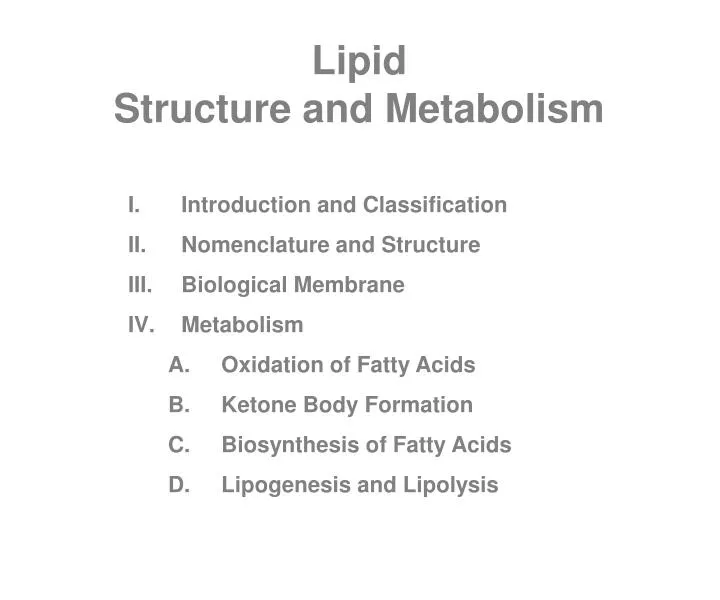
Lipid Structure and Metabolism. Introduction and Classification Nomenclature and Structure Biological Membrane Metabolism Oxidation of Fatty Acids Ketone Body Formation Biosynthesis of Fatty Acids Lipogenesis and Lipolysis. Introduction.

E N D
Lipid Structure and Metabolism • Introduction and Classification • Nomenclature and Structure • Biological Membrane • Metabolism • Oxidation of Fatty Acids • Ketone Body Formation • Biosynthesis of Fatty Acids • Lipogenesis and Lipolysis
Introduction “Biological molecules that are insoluble in aqueous solutions and soluble in organic solvents, have some relation to fatty acids as esters, and have potentiality of utilization by living organisms are classified as lipids.” • They perform four major physiological functions: • Serve as structural components of biological membranes • Provide energy reserves, predominantly in the form of triacylglycerols • Both lipids and lipid derivatives serve as vitamins and hormones • Lipophilic bile acids aid in lipid solubilization
Classification • Bloor’s Classification • A. Simple lipid - ester of fatty acids with various alcohols • 1. Natural fats and oils (triglycerides) • 2. Waxes • (a) True waxes: cetyl alcohol esters of fatty acids • (b) Cholesterol esters • (c) Vitamin A esters • (d) Vitamin D esters • Compound lipid - esters of fatty acids with alcohol plus other groups • 1. Phospholipids and spingomyelin: contains phosphoric acid and often a nitrogenous base • 2. Spingolipids (also include glycolipids and cerebrosides): contains aminoalcohol spingosine, carbohydrate, N-base; glycolipids contains no phosphate • 3. Sulfolipids : contains sulfate group • 4. Lipoproteins : lipids attached to plasma/other proteins • 5. Lipopolysaccharides: lipids attached to polysaccharides
Classification cont. • Derived lipids – hydrolytic products of A & B with lipid characters • 1. Saturated & unsaturated fatty acids • 2. Monoglycerides and diglycerides • 3. Alcohols (b-carotenoid ring, e.g., vitamin A, certain carotenoids) • Miscellaneous lipids • 1. Aliphatic hydrocarbons: found in liver fat and certain hydrocarbon found in beeswax and plant waxes • 2. Carotenoids • 3. Squalene : found in shark and mammalian liver and in human sebam; an important intermediate in biosynthesis of cholesterol • 4. Vitamin E and K
Figure 43 Nomenclature and Structure • Fats and oils: • Vegetable oils are triglycerides that are liquid at room temp due to their higher unsaturated or shorter-chain fatty acids • Triglycerides are most abundant natural lipids • Natural fats have D-configuration • Usually R1 and R3 are saturated and R2 is unsaturated • Natural fats are mixture of two or more simple triglycerides
Fatty acids “ A fatty acid may be defined as an acid that occurs in a natural triglyceride and is a mono carboxylic acid ranging in chain length From four carbon to 24 carbon atoms and including , with exceptions, only the even-numbered members of the series ”
Figure 44Some Natural Fatty Acids Hydroxy acids : ricinoleic acid and dihydroxy stearic acid (castor oil) cerebronic acid (C23H46[OH]COOH, 2 – hydroxy tetracosanoic acid) (cerebroside of animal tissues) Cyclic acids: Hydnocarpic and chaunmoogric acids (chaulmoogra oil, used in treatment of leprosy) Linoleic acid, linolenic acid and arachidonic acid are essential fatty acids
Figure 45 Oleic acid is D9; linoleic acid is D9,12, g-linolenic acid is D6,9,12, arachidonic acid is D5,8,11,14. Natural unsaturated fatty acids are all cis isomers. The schematic form of linoleic acid is as follows:
Phospholipids • They are first and foremost structural components of membranes • Serves as emulsifying agents and surface active agents • They are amphipathic molecules • They are of two types : phosphoglycerides and spingomyelins (contains spingosine instead of glycerol) • Ceramide is the core structural unit of spingolipids which is a fatty acid amide derivative of spingosine
Steroids • Contains cyclopentanoperhydrophenanthrene ring • It is the nonsaponifiable fraction of lipids • Main constituent of animal tissues and abundant in brain, nerve tissue and glandular tissue • Chief component of gallstones • Normal blood level is 200 mg/ml a portion of which is plasma protein bound
Key Words of Today’s Lecture Lipid Triglycerol Fatty Acid Phospholipid Spingolipid Cholesterol
Eicosanoids • Extremely powerful hormone like molecules but are not hormones rather autocrine regulators • Derived from arachidonic acid which is synthesized from linoleic acid by adding a two carbon unit and inserting two additional double bonds • Phospholipase A2 release arachidonic acid from membrane phopholipid to initiate eicosanoid synthesis • Prostaglandins , thromboxanes and leukotrienes are the three types of eicosanoids
Key Features of Eicosanoids • Prostaglandins contains a cyclopentane ring with hydroxy group at C - 11 and C - 15 (pain, fever, ovulation, uterine contraction, gastric secretion inhibition) • Thromboxanes possess a cyclic ether in their structures; TxA2 is the most prominent member of this group and is primarily produced by platelet (clotting) • Leukotrines are hydroxy derivatives possessing conjugated trienes ; early discovery was in leukocites. LTC4, LTD4 and LTE4 are “ slow - releasing substance of anaphylaxis ” ( SRS - A ) , cause fluid leakage from blood vessels to inflamed area. LTB4 is a potent chemotactic agent
Figure 52Biological Actions of Selected Eicosanoid Molecules
Lipoproteins “Proteins covalently linked to lipid found in the blood plasma” Key concepts Plasma lipoproteins transport lipid through the bloodstream. On the basis of density, lipoproteins are classified into four major classes: • Chylomicrons: Large lipoproteins of extremely low density; transport dietary triglycerols and cholesteryl esters from intestine to the tissues (muscle/ adipose) • VLDL (0.95-1.006 g/cc) : synthesized in the liver ; transport lipids to tissues; coverted to LDL with depletion of triglycerol, apoproteins and phospholipids • LDL (1.006 - 1.063 g/cc): carry cholesterol to tissues; engulfed by cells after binding to LDL receptors • HDL (1.063 - 1.210 g/cc ): produced in liver; scavenge cholesterol from cell membrane as cholesteryl ester which is transported to liver from where the excess cholesterol is disposed of as bile acids Atherosceloresis is A chronic disease in which soft masses called atheromas, accumulate on the inside of arteries”
Figure 53 General Structure of Plasma Lipoproteins
Figure 54A Summary of the Relative Amounts of Cholesterol, Cholesteryl Ester, Phospholipid, and Protein in Four major Classes of Plasma Lipoproteins
Membrane Lipids According to the fluid mosaic medel, membrane is a lipid bilayer; proteins float within the bilayer and determines the membrane’s biological function Chemical Composition of Some Cell Membranes
Figure 55 Structure of Lipid Bilayer
Figure 56 Membrane Structure Majority of membrane lipids are phospholipids which are largely responsible for following biological events of membranes: • Membrane fluidity • Selective permeability • Self-sealing capability • Asymmetry
Key Words of Today’s Lecture Eicosanoids Prostaglandins Thromboxanes Leukotrienes Plasma Lipoproteins Membrane Lipids
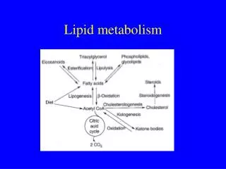
Metabolism The hydrophobic and highly reduced structure of triglycerols allows them to serve as a compact and rich source of energy (e.g., in average U.S. diet, 30 – 40% of calories are provided by fat). The metabolism of lipids include the degradation and synthesis and regulation of these processes. A major emphasis is placed on the role of the central metabolite in lipid metabolism: acetyl-CoA.
Figure 57Oxidation 1. ACTIVATION Thiokinases also known as Acyl-CoA ligase activate the fatty acids to fatty acyl-CoA Thiophorases activate by interconversion
2. Acyl-CoA Translocation: Role of Carnitine Acyl - CoA ligase is found in the outer mitochondrial membrane. Mitochondrial inner membrane is impermeable to most acyl-CoA and carnitine is used to transport these acyl groups as follows: • Each acyl - CoA is converted to acylcarnitine; carnitine acyltransferase I catalyze this reaction • A carrier protein within the mitochondrial inner membrane transfers acylcarnitine into the mitochondrial matrix • Acyl - CoA is regenerated by carnitine acyltransferase II • Carnitine is transported back into the inner membrane by carrier protein and the cycle goes on
Stoichiometry for Palmitic Acid Oxidation Each FADH2 yield 1.5 ATP and NADH 2.5 ATP during electron transport and oxidative phosphorylation, acetyl CoA yields 10ATP thus a total of 108ATP of which 2 ATP is utilized in conversion of palmitic acid to Palmityl-CoA, thus 106 molecules ATP per molecule of palmitic acid is synthesized
Oxidation of Unsaturated Fatty Acids • Similar pathway as above • Usually natural unsaturated fatty acids have cis configuration, which is transformed to trans by the action of an isomerase, as enoyl hydrates acts only on trans double bonds. • An epimerase is needed to form L-isomer as D-isomer of 3-hydroxy fatty acyl-CoA is formed when enoyl hydrases acts on D2,3 unsaturated fatty acid
Figure 59 Oxidation of Odd-chain Fatty AcidsConversion of Propionyl-CoA to Succinyl-CoA End product of b-oxidation of an odd-number fatty acid is acetyl-CoA and a propionyl-CoA. The propionyl-CoA is then converted to succinyl-CoA, a citric acid cycle intermediate
Ketone Bodies Most of the acetyl-CoA product during fatty acid oxidation is utilized by the citric acid cycle or in isoprenoid synthesis. In a process called ketogenesis, acetyl–CoA molecules are used to synthesize acetoacetate, b-hydroxy butyrate and acetone, a group of molecules called the ketone bodies Ketone body formation occurs within mitochondria Ketone bodies are used to generate energy by several Tissues, e.g., cardiac and skeletal muscle and brain
Figure 61Conversion of Ketone Bodies to Acetyl-CoA
Ketosis In normal metabolic pathway, acetoacetate and b-hydroxybutyrate are the ketone bodies which are converted to acetyl - CoA. However, during starvation and in uncontrolled diabetes, conc. of acetoactate is very high and supply of oxaloacetate (a TCA component) is insufficient, thus acetoacetate spontaneously decarboxylated to acetone - KETOSIS • A 4-carbon acid (oxaloacetate) is needed to react with excess acetyl-CoA and form citrate • When OAA is not available excess acetyl - CoA in liver are condensed to form ketone bodies • OAA is limited during scarcity of glucose for glycolysis. In starvation and diabetes, glycogen is broken down. Fatty acids of fat depots are metabolized to supply ATP needs producing excess of the ketone bodies
Key Words of Today’s Lecture b-Oxidation Carnitine Acyl-CoA & acetyl-CoA Propionyl-CoA & Succinyl-CoA Ketone Bodies Ketosis
The oxidation of fatty acids requires the following five enzymes: • 1. Fatty acyl-CoA dehydrogenase, 2. beta-hydroxyacyl-CoA dehydrogenase, 3. beta-ketothiolase, 4. Enoyl hydrase, 5. Thiokinase. Which is the order of the enzyme involved? • A. 1, 2, 3, 4, 5 • B. 2, 3, 4, 5, 1 • C. 5, 2, 4, 1, 3 • D 5, 1, 4, 2, 3 • E. 3, 2, 4, 1, 5 • Which is responsible for the transporting of fatty acid across the mitochondrial inner membrane? • NADH • Plasmin • Cyctochrome Q • Carnitine • Ketosis is ascribed in part to: • A. Slowdown in fat metabolism • B. An insufficient intermediates of TCA cycle • C. An underproduction of acetyl-SCoA • A vigorous protein synthesis • An inhibition of glycogen synthesis • Beta-oxidation of octanoic acid in mitochondria yields: • A. 1 FADH2, 1 NADH and 1 acetyl-CoA • B. 2 FADH2, 2 NADH and 2 acetyl-CoA • C. 2 FADH2, 2 NADH and 3 acetyl-CoA • D. 3 FADH2, 3 NADH and 3 acetyl-CoA • E. 3 FADH2, 3 NADH and 4 acetyl-CoA
Which of the following is not true regarding the oxidation of the mole of palmitate (16) by the β-oxidation pathway? • A. 8 moles of acetyl-CoA are formed • B. 1 mole of the ATP is needed • C. 8 moles of FADH2 are formed • D. The reactions occur in the mitochondria • E. AMP and PP are formed • A fatty acid with an odd number of carbons will enter the citric acid cycle as acetyl-CoA and: • A. α-ketoglutarate • B. Malate • C. Succinyl-CoA • D. Citrate • E. Butyrate • Which of the following statements apply to the β-oxidation of fatty acids? • A. The process takes place in the cytosol of mammalian cells. • B. Carbon atoms are removed from the acyl chain one at a time. • C. Before oxidation, fatty acids must be converted to their CoA derivatives. • D. NADP+ is the electron acceptor. • Carnitine is: • A. One of the amino acids commonly found in protein. • B. Present only in carnivorous animals. • C. Essential for intracellular transport of fatty acids. • D. An essential cofactor for the citric acid cycle. • E. A 15-carbon fatty acid.
Formation of Malonyl-SCoA This is considered as activation step as the breaking of the CO2 bond of malonyl-SCoA releases lot of energy that “drives” the reaction forward
Figure 63Diagrammatic View of Fatty Acid Biosynthesis
Lipogenesis and Lipolysis • Fatty acid may be converted to triacylglycerol, degradated to generate energy, or utilized in membrane synthesis • When serum glucose level is high after meal, insuline promotes triacylglycerol synthesis by facilitating the transport of glucose into adipocytes - lipogenesis • Adiposites can not synthesize triacylglycerol when glucose level is low (between meal), when several hormones stimulate the hydrolysis of triacyl-glycerol within adipose tissue to form glycerol and fatty acids – lipolysis.
Figure 65Biosynthesis of Phosphatidic Acid and Triglyceride
Lipolysis Lipolysis occur during fasting, vigorous exercise, and in response to stress. Binding of several hormones (e.g., glucagon and epinephrine) to specific adipocyte plasma membrane receptors initiates a reaction sequence similar to the activation of glycogen phosphorylase. As a result of cAMP synthesis, the enzyme triacylglycerol lipase (hormone-sensitive lipase) is activated when it is phosphorylated by protein kinase which is activated by cAMP