Download
1 / 22
220 likes | 332 Views
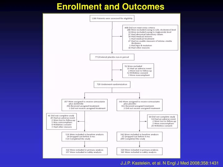
Enrollment and Outcomes. J.J.P. Kastelein, et al. N Engl J Med 2008;358:1431. Baseline Characteristics of the Patients*. J.J.P. Kastelein, et al. N Engl J Med 2008;358:1431.

E N D
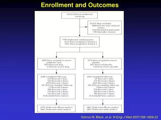
Enrollment and Outcomes J.J.P. Kastelein, et al. N Engl J Med 2008;358:1431
Baseline Characteristics of the Patients* J.J.P. Kastelein, et al. N Engl J Med 2008;358:1431
Levels of Lipids, Lipoproteins, Sterols, and C-Reactive Protein at Baseline and after 24 Months of Treatment, with Changes from Baseline* - Part I J.J.P. Kastelein, et al. N Engl J Med 2008;358:1431
Levels of Lipids, Lipoproteins, Sterols, and C-Reactive Protein at Baseline and after 24 Months of Treatment, with Changes from Baseline* - Part II J.J.P. Kastelein, et al. N Engl J Med 2008;358:1431
Effects of Simvastatin and Combined Therapy with Simvastatin plus Ezetimibe on Levels of Cholesterol and Triglycerides J.J.P. Kastelein, et al. N Engl J Med 2008;358:1431
Measures of Intima–Media Thickness in Carotid and Femoral Arteries at Baseline and at 24 Months and Changes from Baseline* J.J.P. Kastelein, et al. N Engl J Med 2008;358:1431
Mean (±SE) Intima–Media Thickness of the Carotid Artery during 24 Months of Therapy J.J.P. Kastelein, et al. N Engl J Med 2008;358:1431
Carotid arterial segments. The common carotid (defined as the segment 1cm proximal to the dilatation), carotid bulb (between the dilatation and the flow divider) and the internal carotid (1cm distal from the flow divider) arterial segments. Carotids are scanned bilaterally J.J.P. Kastelein, et al. N Engl J Med 2008;358:1431Supplementary Appendix
Femoral arterial segment. The common femoral (defined as the segment 1cm proximal to the dilatation) arterial segment. Femorals are scanned bilaterally J.J.P. Kastelein, et al. N Engl J Med 2008;358:1431Supplementary Appendix
B-mode ultrasound still image of the common carotid arterial wall segment and its standard settings as used in the imaging trial proptocol. The arrow in the far wall indicates the carotid flow divider. The image is saved as a TIFF-file J.J.P. Kastelein, et al. N Engl J Med 2008;358:1431Supplementary Appendix
M-mode images are created in the distal common carotid artery J.J.P. Kastelein, et al. N Engl J Med 2008;358:1431Supplementary Appendix
The near and far walls are visualized in the 1/3 B-mode; the movement in time is depicted in the 2/3 M-mode image. The 1/3-2/3 image is saved as a TIFF-file of the same format as the Bmode image J.J.P. Kastelein, et al. N Engl J Med 2008;358:1431Supplementary Appendix
Image analysis of a the region of interest of a B-mode image, using the eTrack program. The image analyst (or ‘reader’) indicates the lumen-intima and media-adventitia interfaces with cross-hairs using an interactive cursor. The cross-hairs are splined J.J.P. Kastelein, et al. N Engl J Med 2008;358:1431Supplementary Appendix
Control image as used for B-mode image analysis. The control image represents the ROI of the image. The splined (manual) tracings delineate the lumen-intima and the media adventitia interfaces. The light gray bar on the right side of the control image indicates the videolines that could (homogeneous gray) and could not be (striping) evaluated by the image interpolation function of the image analysis program. In this example, the striping is in the lumen and hence outside the region of interest of and of no influence on the IMT measurement J.J.P. Kastelein, et al. N Engl J Med 2008;358:1431Supplementary Appendix
Image analysis of a the region of interest of an M-mode image, using the eTrack program. At least 2 heartbeats are required for a proper tracing of the lumen and arterial wall stiffness parameters J.J.P. Kastelein, et al. N Engl J Med 2008;358:1431Supplementary Appendix
Control image as used for M-mode image analysis. The (automatic) contour tracings delineate the peri-adventitial – adventitia interface and the intima-lumen interfaces of the near wall, and the lumen-intima and the media-advential interfaces of the far wall. The lumen is measured between the leading edges of the interfaces of the intima-lumen of the near and the lumenintima of the far wall interfaces J.J.P. Kastelein, et al. N Engl J Med 2008;358:1431Supplementary Appendix
Most common Treatment Related Treatment Emergent Adverse Events J.J.P. Kastelein, et al. N Engl J Med 2008;358:1431Supplementary Appendix
Commonly Reported Treatment Emergent Adverse Events J.J.P. Kastelein, et al. N Engl J Med 2008;358:1431Supplementary Appendix
Adverse event leading to withdrawal of consent J.J.P. Kastelein, et al. N Engl J Med 2008;358:1431Supplementary Appendix
Additional secondary endpoints J.J.P. Kastelein, et al. N Engl J Med 2008;358:1431Supplementary Appendix
Prespecified exploratory analyses J.J.P. Kastelein, et al. N Engl J Med 2008;358:1431Supplementary Appendix
Treatment difference in change in cIMT in prespecified subgroups Bars represent (95% confidence interval) J.J.P. Kastelein, et al. N Engl J Med 2008;358:1431Supplementary Appendix