Download
1 / 65
710 likes | 1.33k Views
Bleeding Late in Pregnancy. When the placenta misbehaves Grace Cavallaro MD, FACOG. Objectives. Identify major causes of vaginal bleeding second half of pregnancy Describe a systematic approach to identify the cause of bleeding Describe specific treatment options based on diagnosis.

E N D
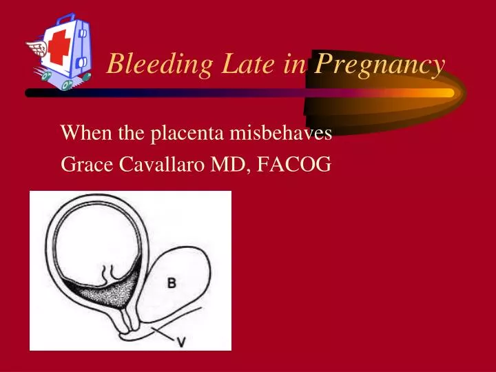
Bleeding Late in Pregnancy When the placenta misbehaves Grace Cavallaro MD, FACOG
Objectives • Identify major causes of vaginal bleeding second half of pregnancy • Describe a systematic approach to identify the cause of bleeding • Describe specific treatment options based on diagnosis
Causes of Late Pregnancy Bleeding • Placenta Previa • Abruption • Ruptured Vasa Previa • Uterine Scar Disruption • Cervical Polyp • Bloody Show • Cervicitis • Vaginal Trauma • Cervical Cancer Life Threatening*
Prevalence of Placenta Previa • Occurs in 1/200 pregnancies that reach 3rd trimester • Low-lying placenta seen in 50% of ultrasound scans at 16-20 weeks • 90% will have normal implantation when scan repeated @ >30 weeks • No proven benefit to routine screening ultrasound for this diagnosis.
Risk factors for previa • Previous Cesarean Sections • Previous Uterine Instrumentation • High Parity • Advancing Maternal Age • Women over 40 have a RR of 9.0 • Smoking • Multiple Gestation
Morbidity with Placenta Previa • Maternal Hemorrhage • Operative Delivery Complications • Transfusion • Placenta accreta, increta or percreta • Prematurity
Placenta Migration • Migration means the dynamic relationship between the placenta and the internal os • Trophotropism vs elongating lower uterine segment!
Previous C-sections and Previas Anath ObGyn 1996
Patient History - Placenta Previa • Painless Bleeding* • 2nd or 3rd trimester, or at term • Often following intercourse • May have preterm contractions* • Sentinel Bleed • From large central previa • @ 26-28 weeks gestation
Physical Exam-Placenta Previa • Vital Signs • Assess Fundal Height • Fetal Lie • Estimated Fetal Weight (Leopold) • Presence of fetal heart tones • Gentle Speculum Exam • No digital exam unless placental location known
Laboratory - Placenta Previa • Hematocrit or complete blood count • Blood Type and Rh • Coagulation tests • (While waiting - serum clot tube taped to the wall)
Ultrasound - Placenta Previa • Can confirm diagnosis • Full bladder can create false appearance of anterior previa • Presenting part may overshadow posterior previa • Transvaginal scan can locate placental edge and internal os
The Placenta’s Ultrasound Appearance Echodense placental tissue Echolucent myometrial Area rich in blood supply
Vagina and Cervix meet at 90 degrees Careful insertion of the vaginal probe midway into the vagina will image the LUS and the cervical os
Posterior Previa Transvaginal Scan Posterior Placenta Previa
False Previa c Full bladder No Previa Lower placental border
False Previa - Overdistended Bladder Bladder Cervical canal c
Placental Edge by U/S and Route of Delivery • >2 cm os - placenta edge = safe for vaginal delivery • <1cm os - placenta edge - Cesarean delivery • 1-2 cm = may be able to deliver vaginal • Dawson et al Jultrasound Medicine 1996
Ultrasound’s Role • Previa = usually definitive except in very low lying posterior placentas in the obese patient • Abruption - definitive diagnosis is not possible • Transvaginal Scanning is safe in the bleeding patient
Clinical Signs and Symptoms • Painless Bleeding = Previa • Painful Bleeding = Abruption • Painless Fetal Bleeding = Vasa Previa
Initial management • 1) ABC’s • Amount of bleeding noted is unreliable • 2) Fetal Well Being • 3) No Vaginal Exams • Until you know where the placenta is! • 4) Ultrasound
Fetal/Neonatal Considerations • Gestational Age of Fetus dictates local of care • SGA/Prematurity are major problems • Communication with consultants is key!
Cesarean Sections and Previas Category C • Pre-op Scan • Patients with Previas undergoing C-Section • Bleed More • Require More Blood Transfusion • Require More C-Hysterectomies • Placenta accreta may accompany 10% • Bladder invasion may be associated with • DIC and massive hemorrhage
Treatment Placenta Previa • With no active bleeding • Expectant management • No intercourse, digital exam • Rescan after 30 weeks • With late pregnancy bleeding • Assess overall status, circulatory stability • Full dose Rhogam if Rh - • Consider maternal transfer if premature • May need corticosteroids, tocolysis, amniocentesis Category B
Expectant Management • May discharge home if stable after 72 hours of inpatient observation. • Reduces stay in hospital by average of 14 days. • No increase in • Hemorrhage • Need for transfusion • Poor maternal or neonatal outcomes
Tocolytics in Placenta Previa • Greatest morbidity and mortlity related to prematurity. • Tocolytics can add an additional 11 days to pregnancy. • Allows for administration of corticosteroids • No increase in maternal or fetal complications • Increase birth weights average of 320 grams
Appropriate only in marginal (anterior) previa with vertex presentation Palpation of placental edge and fetal head with set up for immediate surgery Cesarean delivery under regional anesthesia if Complete previa Fetal head not engaged Non-Reassuring tracing Brisk or Persistant bleeding Mature fetus Double Set-up Exam: digital exam in OR with ability to do immediate CD Category D
Placental Abruption • Premature separation of placenta from uterine wall • Partial or Complete • “Marginal sinus separation” or “marginal sinus rupture” • Bleeding, but abnormal implantation or abruption never established
Epidemiology of Abruption* • Occurs in 1-2% of all pregnancies • Risk Factors • Hypertensive diseases of pregnancy • Smoking or substance abuse* • Trauma* • Overdistension of the Uterus* • History of Previous Abruption* • Unexplained elevation of MSAFP • Placental insufficiency • Maternal Thrombophilia/Metabolic abnormalities
Abruptions and Trauma • Can occur with blunt abdominal trauma and rapid deceleration without direct trauma • Complications include prematurity, growth restriction and stillbirth • Fetal evaluation after trauma • Increased use of FHR monitoring may decrease mortality Category C
Bleeding from Abruption • Externalized hemorrhage • Bloody amniotic fluid • Retroplacental clot • 20% occult • “uteroplacental apoplexy or Couvelaire uterus • Look for consumptive coagulopathy
Cigarette Smoking as Risk factor • Nova Scotia Registry of 87, 184 pregnancies • 33% smoked • 2.05 Relative Risk of Abruption • 1.75 Relative Risk of Previa • No dose effect noted • Anath AmJ of Epidemiology 1996
Cocaine/Metamphetamine • Associated with • chorionic villous hemorrhage • Villous edema • Even in the absence of clinical abruption placenta
Patient History: Abruption • Pain = hallmark symptom* • Varies from mild cramping to severe pain • Back Pain - think posterior abruption • Bleeding • May not reflect amount of blood loss* • Differentiate from exuberant bloody show • Trauma • Other risk factors (e/g hypertension/drugs) • Membrane rupture
Physical Exam- Abruption • Signs of circulatory instability • Mild tachycardia normal • Signs and symptoms of shock represent > 30% blood loss • Maternal abdomen • Fundal height • Leopold’s:estimated fetal weight, fetal lie • Location of tenderness • Tetanic contractions
Ultrasound Abruption • Abruption is a clinical diagnosis!* • Placental location and appearance • Retroplacental echolucency • Abnormal thickening of placenta • “Torn” edge of placenta • Fetal lie • Estimated fetal weight
Placental Abruption Hemorrhage isoechoic with placenta Hematoma retroplacental
Abruption - Retroplacental Hematoma 7 days later Retro placental hematoma day1
False Abruption? Contraction Mimicking Abruption Contraction No Contraction 30 minutes later
Placenta Lakes Subchorionic Placental Lake Doppler revealing flow through the lake
Laboratory-Abruption • Complete blood count • Type and Rh • Coagulation tests + “Clot test” • Kleihauer-Betke test not diagnostic, but useful to determine Rhogam dose • Pre-eclampsia labs, if indicated • Consider urine drug screen
Treatment-Grade II Abruption • Assess fetal and maternal stability • Amniotomy • IUPC to detect elevated uterine tone • Expeditious operative or vaginal delivery • Maintain urine output > 30 cc/hr and hemotocrit > 30% • Prepare for neonatal resuscitation Category C
Treatment - Grade III Abruption • Assess mother for hemodynamic and coagulation status • Vigorous replacement of fluid and blood products • Vaginal delivery preferred, unless severe hemorrhage Category C