Download
1 / 103
1.03k likes | 1.22k Views
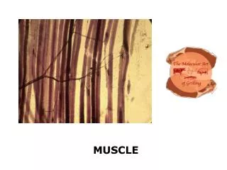
MUSCLE I. MUSCLE TISSUE Unicellular organisms have developed cytoplasmic specializations responsible for motility (cilia and flagella). Multicellular organisms have adapted a class of mesodermally-derived cells specialized for contractile ability.

E N D
MUSCLE TISSUE • Unicellular organisms have developed cytoplasmic specializations responsible for motility (cilia and flagella). • Multicellular organisms have adapted a class of mesodermally-derived cells specialized for contractile ability.
Muscle tissue is one of the 4 basic tissue types The general structure of muscle is a striking example of how “ FORM FOLLOWS FUNCTION ”
Composed of two basic elements: • Muscle cells: (fibers) • develop contractile force through shortening of the cell • Connective Tissue: • Provides a framework against which the contractile forces of muscle cells can be exerted. • Surrounds and support aggregates of muscle fibers providing nutrients, oxygen and nerve innervation. • Muscle tissue is provided with a rich complement of Bloodvessels & Nerves
Functions of Muscle: 1. Movement External - skeletal (movement of your 206 bones & joints) Internal - cardiac = blood thru circulatory system - smooth =movement thru tubular systems (food thru GI tract, Blood Pressure) 2. Posture - tonic control of axial muscles against gravity (isometric contraction) 3. Heat - generation of body heat, 85% (shivering= oscillating contraction/relaxation)
Structural Requirements of Muscle 1. Generation of Tension: (shortening, 2/3 of its length) 2. Attachment: (connective tissue) 3. Control of Contraction: - on/off - magnitude - precise controlled movement - communication - coordinated contraction of all cells - coordinated nerve muscle signalling 4. Repair: after damage or injury - cell differentiation - mitotic division
General Organization & Nomenclature: Muscle: many muscle cell groups (fascicles) surrounded by a sheath of dense irregular CT epimysium (type I collagen) Fascicle: groups of muscle fibers (cells) - surrounded by septa of CT from the epimysium - perimysium (type I collagen) Fiber: an individual muscle cell (long cylindrical cells) - surrounded by loose CT - endomysium and a basal lamina (type I, IV collagen). Muscle cell = Muscle fiber = Myocyte
Within each individual muscle cell: Myofibrils: - structural units within the muscle cell sarcoplasm: - composed bundles of two types of filaments = myofilaments (hundreds / myofibril) Myofibrils are held in precise alignment by intermediate filaments (desmin, vimentin, dystrophin) Myofilaments: - “ thin (actin) and “thick” (myosin) protein chains. The precise alignment of these filaments gives the appearance of striations of the LM level
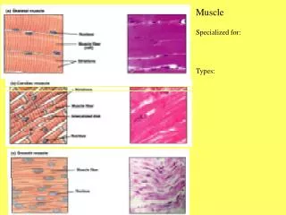
3 types of muscle: Skeletal - Cardiac - Smooth Muscle Classification Schemes: 1. Structural organization: - Presence or absence of striations 2. Control of Contraction: - Voluntary or involuntary control
FEATURE SKELETAL CARDIAC SMOOTH CELL SHAPE CYLINDRICAL BRANCHED FIBERS FUSIFORM SARCOMERE YES YES NO NUCLEI / CELL (LOCATION) MULTIPLE (PERIPHERAL) 1 (CENTRAL) 1 (CENTRAL) CELL JUNCTIONS NONE INTERCALATED DISK , Gap JX GAP JUNCTIONS Control of CONTRACTION VOLUNTARY “ALL OR NONE” INVOLUNTARY RHYTHMIC, SPONTANEOUS INVOLUNTARY SLOW, FORCEFUL REGENERATION YES (SATELLITE CELLS) NO YES (MITOSIS) MITOTIC DIVISION NO NO YES
Muscle Nomenclature: Individual Muscle Cells (myofibers) 10 - 100um diameter - several millimeters long, multinucleated cells (can be as long as 30 cm) multiple nuclei are located at the periphery of the cytoplasm. The standard cytoplasmic organelles have become highly specialized in these cells and are given different names, usually preceded by the prefix “Myo” or “Sarco”
SKELETAL MUSCLE CELL COMPONENTS: - plasmalemma = sarcolemma (specialized invaginations, T - tubules) - cytoplasm = sarcoplasm - endoplasmic reticulum = sarcoplasmic reticulum (very extensive- highly specialized) - cytoplasmic filaments = myofilament - Bundles (1-3um diameter) = myofibrils - Individual = thin/thick filaments - Mitochondria = sarcosomes (usually encircle the myfibrils near the I-band)
MUSCLE FIBER TYPES: Skeletal muscle fibers contain the protein Myoglobin - similar to hemoglobin in blood in that it serves as an oxygen donor for the metabolic machinery of the cell. Skeletal muscle fibers can be divided into 3 types according to their Myoglobin content RED – WHITE - INTERMEDIATE
REDWHITEINTERMEDIATE (Type I) (Type II B) (Type II A) Small Large Intermediate myoglobin myoglobin intermediate glycogen glycogen glycogen mitochondria mitochondria mitochondria Slow Very Fast Moderately Fast contraction contraction contraction long acting rapidly fatigued intermediate Primarily Primarily Both Oxidative Glycolytic aerobic ATP anaerobic ATP aerobic & anaerobic production production production
Bands in Striated Muscle - In the light microscope muscle cells cut in longitudinal section exhibit a characteristic pattern or banding or “striations”. - This reflects the ultrastructural organization of the myofibrils which are arrayed in precise and regular alignment within the sarcoplasm.
A-Band: dark staining band I-Band: light staining band Z-line: dark line which bisects the I band This series of alternating light and dark bands is the light microscopic appearance of the basic contractile unit of the muscle cell the “sarcomere”.
THE SARCOMERE • composed of the region from one Z line to another • approx 2.5um 1.5um = A- band • 1.0um = I-band. • The sarcoplasm of an individual muscle cell is filled with long cylindrical bundles called myofibrils about (1-2um in diameter) - • aligned parallel to the long axis of the muscle fiber.
Each myofibril exhibits the same banding pattern as the muscle cell. The precise lateral alignment of myofibrils within the muscle cells that gives the appearance that the striations run across the entire width of a cell. If we examine a single myofibril at the EM level it is possible to distinguish two additional bands of the sarcomere. - H-Zone: light staining region in the center of the A-band - M-Line: dark staining line in the center of the H-Zone
BANDS OF THE SARCOMERE: A-BAND = Region of overlapping thick and thin filaments I-BAND = Thin filaments only H-BAND = Thick filaments only M-Line = Thick filaments and crossbridges Z-Line = alternately spaced thin filaments from adjoining sarcomeres anchored by protein crossbridges
THE Z-LINE 3 Major Proteins 1. Z-Line Protein Anchoring protein 2. Alpha Actinin (binding protein for actin) 3. Cap-Z (caps the (+) ends of the thin filaments)
THIN FILAMENTS(and associated proteins) • Actin (polarized having a plus + (attached) and minus (free) end • – helical dimer • 2. Tropomyosin (regulates availability of myosin binding sites • 3. Troponin (Ca+ regulation of contraction) • 4. Nebulin (anchored at Z-line, runs the length of each actin filament) • 5. Tropomodulin (cap protein for (-) end of actin filaments)
THIN FILAMENTS:Approx. 1.0um long, 6-7 nm diam -filamentous actin (F-actin) formed from two strands of globular actin (G-actin) arranged in a double helix. - Strands of the protein tropomyosin are associated with the actin double helix in the groove between the two F - actin strands. - This is the location of the myosin binding site on the actin molecule - At rest the structural conformation of the tropomyosin strands is such that tropomyosin covers these myosin binding sites on the actin filament.
A globular protein troponin attaches to the tropomysin strands at regular intervals. Troponin has 3 subunits: - Tn tropomyocin binds to the tropomyosin strand - Tn inhibit inhibits (regulates the covering of ) the myosin binding site on actin filaments - Tn calcium binding site for calcium during the initiation of muscle contraction
THICK FILAMENTSand associated proteins • 1. Myosin: major structural protein (dimer) • M-line Proteins: • - myomesin holds myosin bundles apart to • allow for overlap of thin filaments • - creatine kinase catalyzes the conversion of • ADP > ATP • 3. C-protein: binds myosin strands together into a thick filament • 4. Titin: Anchors the tail regions of the myosin dimers
MYOSIN (THICK) FILAMENTS approx 1.5um long, 12-15nm diam - composed primarily of multiple strands of a myosin dimer held together by a structural protein termed C-Protein. Each myosin monomer is composed 3 regions: which together take a “golf club” like shape - a short head region (heavy meromyosin) - a short neck region (heavy meromyosin) - a longer tail region (light meromyosin)
The head/neck region of myosin contains 3 important components 1. Actin binding site 2. ATP binding site 3. Flexible region capable of conformational change Two myosin light chain proteins which wrap around the myosin neck * essential light chain * regulatory light chain The tail regions of the myosin are anchored to an extremely large the protein TITAN, which anchors to the Z-line at one end and the tail region of the thick filaments at the other.
SUMMARY OF THE PROTEIN STRUCTURE OF THE SACOMERE
T-tubules and the Sarcoplasmic Reticulum: Muscle contraction predominently depends on 2 physiological events: 1. Depolarization of the muscle cell membrane 2. The release of Ca++ ions from intracellular stores within the muscle cell sarcoplasmic reticulum Muscle cells have evolved specialization of both intracellular and plasma membranes to assist in these processes
Sarcoplasmic Reticulum: - very extensive within a single muscle cell - forms large bulbous terminal cisternae at it’s terminal ends near the junction of the A and I bands - The SR completely surrounds each myofibril - serve as a major reservoir for Ca++ ions during muscle contraction
T-tubules: To effect a more uniform and rapid distribution of membrane depolarization the sarcolemma has evolved a system of invaginating tubules called T-tubules. - deep invaginations of the muscle cell sarcolemma - T-tubules invaginate at the A / I junction and surround this region at each myofibril - make it possible for membrane depolarization to be instantaneously distributed to all myofibrils T-tubules are flanked by a pair of terminal cisternae of the sarcoplasmic reticulum and the three structure are collectively termed “triad”.
Sliding Filament “Theory” of Muscle Contraction: During contraction of a muscle fiber 1. The size of the A-band is unchanged 2. The I-band and H-zone decrease in size The contractile mechanism proposed to account for this is called the “sliding filament theory” of muscle contraction.
The model proposes that during contraction the • thick and thin filaments of a sarcomere slide past one • another causing a: • Shortening of the I-band - due to increased overlap • of thick and thin filaments (reducing the H zone) • The sliding motion is due to the binding of the • myosin head to the actin filaments resulting in a • conformational change in the myosin. • In resting muscle, myosin and actin filaments cannot interact because the binding sites for the myosin head on the actin filaments are blocked by the troponin-tropomyosin complex.
Contraction of a single muscle cell is an all-or- none event, but contraction typically occurs in a smooth precise fashion. • This is due to the unique 3- dimensional arrangement of myofilaments: • interdigitation of thin and thick filaments is not 1:1
A transverse EM view of the sarcomere shows: • Each actin filament is associated with 3 adjacent myosin filaments. • Each myosin filament contacts 6 adjacent actin filaments
5 major steps in muscle contraction: 1. Generation of a nerve impulse (action potential) along an afferent nerve fiber terminating on a muscle cell. 2. Synaptic transmission of the afferent impulse and muscle cell depolarization. 3. Release of Ca++ from the SR. 4. Unmasking of myosin binding sites on actin filaments. 5. Binding of myosin to the actin filament and the “power stroke”.
Step 1. Generation of a nerve impulse • (action potential) along an afferent nerve fiber terminating on a muscle cell. • The initial signal for muscle contraction arises from a nerve impulse generated along the myelinated axons of somatic motor neurons in the central nervous system. • These neurons are located in the ventral horn of the spinal cord.
The terminal ends of their motor axons • end in a specialized contact on muscle • cells termed a “neuromuscular/myoneural • junction”. • A given motor axon can branch at its • tip to contact many individual muscle • cells. • A motor axon and all of the muscle • cells that it contacts are termed a • “motor unit”. • The number of cells in the motor unit determine the precision of the muscle movement.
The myoneural junction and the muscle • synapse: • The point of contact forms a specialized • cell-cell junction • The membranes of the motor axon fan out to • form a specialized contact = “motor • endplate”. • The motor endplate and the muscle cell • sarcolemma become closely apposed in a • shallow invagination of the sarcolemma - • the “primary synaptic cleft”.
The edges of the synaptic cleft are marked by smaller infoldings of the sarcolemma “secondary synaptic clefts” (junctional folds) • which increase the total surface area of the • nerve/muscle contact. • The terminal ends of the motor axons contain numerous intracellular vesicles filled with the neurotransmitter acetylcholine. • The sacolemmal membrane adjacent to the • synaptic cleft contains specialized receptors • for acetylcholine.
STEP 2. Synaptic transmission / muscle depolarization Release of acetylcholine into the synaptic cleft from vesicles in the motor axon terminal results in: * Activation of acetylcholine receptors on the sarcolemma * Opening of ligand-gated Na+ channels in the sarcolemma resulting in initial depolarization of the sarcolemma * Opening of voltage-gated Na+ channels in the sarcolemma resulting in widespread depolarization of the muscle cell
Excitation / Contraction Coupling
Step 3. Release of Ca++ from the SR • (Classic Story) • Depolarization of the sarcolemma stimulates the formation of inositol tri- phosphate (IP3). • IP3 percolates in the sarcoplasm in the region of the SR and binds to an IP3- sensitive Ca++ channel in the terminal cisternae of the SR membrane. • (but see also Dihydropyridine model) • Ca++ is released through these channels and the intracellular levels of Ca++ rise • (10 -7 M -- 3 x 10 -5 M)
(What Really Happens ?) • Depolarization of the sarcolemma activates a dihydropyridine receptor • (DHPR) in the sarcolemma • The DHPR is mechanically coupled ryanodine receptors which are calcium • channels inserted into the SR membrane • Ca++ is released through these channels and the intracellular levels of Ca++ rise • (10 -7 M -- 3 x 10 -5 M)
STEP 4. Unmasking of myosin binding sites • on thin filaments • The increased intracellular Ca++ binds to the Tnc subunit of the troponin complex on the actin filaments. • Binding of Ca++ to the troponin/tropomoysin complex results in a lateral rotation of the tropomyosin strand, driving it deeper into the groove between the two actin helices. • The rotation of the tropomycin uncovers the myosin binding site on the actin filament.
STEP 5. Binding of myosin to the actin • filament and the “power stroke” • With it’s binding site uncovered, myosin is now free to bind to it’s binding site on the actin filaments • At this point ADP + Pi are bound to the head region of the myosin light chain • Unmasking of the myosin binding site by Ca++ allows the myosin to extend out and bind to the actin - this releases the Pi • ADP is then released from the myosin head and the myosin neck flexes = the “power stroke”
The flexion of many myosin heads, ratchets the actin filament along the myosin (power stroke) and the sarcomere shortens. the “power stroke” is estimated at 11nm * the cumulative sequential power strokes of many myosin heads = full muscle cell contraction This cycles is repeated at a rate of 10-50 strokes/second providing: 1. the critical level of intracellular Ca+ is maintained and 2. ATP is available as energy source
THE RIGOR COMPLEX: • Following the power stroke, the myosin remains attached to the actin filament and the • myosin light chains are in their flexed • position • This configuration is known as the “rigor complex” • The rigor complex is maintained indefinitely until: • - ATP binds to the head region of the myosin • - ATP is thus required to break the rigor complex
Muscle Relaxation: • Myofilament attachments release and • sarcomere returns to its resting length • Muscle relaxation is not a passive • process from the standpoint of • bioenergetics. • Nearly 50% of the ATP involved in the • contraction / relaxation cycle is • utilized during relaxation.
Muscle relaxation is a 2 step process: 1. Transport of Ca++ from the sarcoplasm back into the lateral cisternae of the SR. 2. Detachment of Ca++ from the Tnc subunit of troponin and the de-rotation of the tropomyosin out of the actin grove to again cover the myosin binding site