Download
1 / 33
450 likes | 1.49k Views
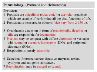
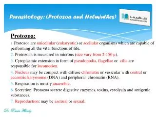
Parasitology: (Protozoa and Helminthes) . Protozoa: Protozoa are unicellular ( eukaryotic ) or acellular organisms which are capable of performing all the vital functions of life. Protozoan is measured in microns ( size vary from 2-150 µ ).

E N D
Parasitology: (Protozoa and Helminthes) Protozoa: Protozoa are unicellular (eukaryotic) or acellular organisms which are capable of performing all the vital functions of life. Protozoan is measured in microns (size vary from 2-150 µ). Cytoplasmic extension in form of pseudopodia, flagellae or cilia are responsible for locomotion. Nucleus may be compact with diffuse chromatin or vesicular with central or eccentric karyosome (DNA) and peripheral chromatin (RNA). Respiration is mostly anaerobic. Secretion: Protozoa secrete digestive enzymes, toxins, cytolysin and antigenic substances. Reproduction: may be asexual or sexual. Dr. Rania Alhady
Parasitology: (Protozoa) According to Mode of locomotion, protozoa are classified into the following groups: Class: Rhizopoda: Protozoa move by pseudopodia. Entamoeba histolytica Class: Mastigophora: Protozoa move by flagellae. Gastrointestinal flagellates: Giardia intestinalis Genitourinary flagellates: Trichomonas vaginalis Blood and Tissue flagellates: Trypanosomagambiense, Trypanosoma rhodesiense ,and Leishmania species. Class: Ciliophora: Protozoa move by cilia. Balantidium coli Class: Sporozoa: Protozoa have no motor organelles . Plasmodium species, and Toxoplasma gondii. Dr. Rania Alhady
Parasitology: (Protozoa) Entamoeba histolytica: • A world wide in distribution • More often in tropical countries with poor sanitary conditions • A commensal protozoa when human has a normal immune function. • Invading host tissues and causing amoebiasis when human has a lower immune function Dr. Rania Alhady
Protozoa: Entamoeba histolytica Morphology:- Trophozoite • No regular in shape, 20~60μm in size. • An active-moving trophozoite produce pseudopods (organelle) • A spherical nucleus. Nucleolus in the center. peripheral chromatin • Erythrophagocytosis RBCs Dr. Rania Alhady
Protozoa: Entamoeba histolytica Cyst • Spherical in shape & 10~20μm in diameter. 1~4 nuclei (similar to that of the trophozoite). • Immature cyst (1 or 2 nuclei) has the glycogen vacuole & chromatoid body. • No inclusions disappear In mature cyst (4 nuclei) ---infective stage Dr. Rania Alhady
Protozoa: Entamoeba histolytica Microscopic characteristics of Entamoeba histolytica: Dr. Rania Alhady
Protozoa: Entamoeba histolytica Characteristic of life cycle • Basic model : cysttrophozoite cyst • Parasitic location : large intestine (common) ; intestinal tissue or other tissues (occasional) • Infective stage : mature cyst • Trophozoite in diarrhea or pus ; Cyst in formed feces • Infection : by ingestion of mature cyst Pathologic changes Colonic tissues : flask-shaped ulcers The destruction of trophozoites on mucosa may be shallow and small. While they enter the submucosa, they multiply and spread laterally give rise to extensive destruction. Extraintestinal tissues (liver) : abscess --- Anchovy-sauce type pus.
Protozoa: Entamoeba histolytica Clinical classification 90% persons infected are carriers • Intestinal amoebiasis Acute intestinal amoebiasis -- amoebic dysentery (bloody, mucus-containing diarrhea) + lower abdominal discomfort + tenesmus Chronic intestinal amoebiasis --- dyspepsia + weight loss + asthenia (common) / diarrhea • Extraintestinal amoebiasis Liver : amoebic hepatitis + amoebic liver abscess --- pain in right-upper-quadrant + fever + marked tenderness of liver Lung : amoebic pulmonary abscess --- pain in chest + cough + fever Sometimes,E.h can be carried to other organs. Such as brain, skin and so on.
Protozoa: Entamoeba histolytica Laboratory diagnosis • Fecal examination --- Wet mounts : Trophozoites in diarrhea feces. --- Wet mounts stained with iodine : Cyst in formed feces. • Pus examination --- Trophozoites in aspirate pus from abscesses
Protozoa: Giardia intestinalis Giardia intestinalis:- • Worldwide distribution • higher prevalence in tropical or developing countries (20%) • 1-6% in temperate countries • Most common protozoa in stools: ~200 million cases/yr • Giardiasis: -often asymptomatic OR -acute or chronic diarrhea Fecal-Oral Transmission Factors • Poor personal hygiene: -children (eg, day care centers) - food handlers • Developing countries: -poor sanitation - endemic -travelers diarrhea • Water-borne epidemics • Male homosexuality: oral-anal contact Dr. Rania Alhady
Protozoa: Giardia intestinalis Pathogenesis: • Trophozoite stage induces malabsorption of fats. • Mechanism(s) unknown. Clinical Disease: Diarrhea (steatorrhea) Weight loss Constipation Fatigue Histopathological correlate: Flattened villi Dr. Rania Alhady
Protozoa: Giardia intestinalis Diagnosis: Identify trophozoites and cysts by microscopic examination of stool Cyst Giardia intestinalis trophozoite: Iron – hematoxylin stain. Dr. Rania Alhady
Protozoa: Trichomonas vaginalis Trichomonas vaginalis • Worldwide in distribution • The most common pathogenic protozoan of human in industrialized countries • Transmission is by contact (by sexual intercourse). Sometimes, by indirect contact, such as sharing damp washclothes / swimming clothes. Morphology (trophozoite) • Pear-like (teardrop), 7~32 X 5~12μm • One nucleus and a axostyle projected posterior out of the body. • Undulating membrane on one side (one-third the length of the body). • Basal body on anterior to nucleus and produce 4 anterior flagella and 1 posterior flagellum.
Protozoa: Trichomonas vaginalis Clinical features : • The incubation period is 5~28 days. • In women, vaginitis with purulent discharge is prominent symptom, be accompanied by vulva and cervical lesions, abdominal pain, dysuria. • In men, asymptomatic (common) ; urethritis, epididymitis and prostatitis (occasional) Laboratory diagnosis • Microscopic examination of wet mounts : detect actively motile organisms. • In women, examination should be performed on vaginal and urethral secretions. • In men, anterior urethral or prostatic secretions should be examined.
Protozoa: Trypanosoma Trypanosoma Trypanosomiasis: Diseases • African Trypanosomiasis American Trypanosomiasis • Sleeping sickness Chagas disease • Transmitted by tsetse fly Transmitted by kissing bug • Large and aggressive fly • Painful bites Triatomid insect Dr. Rania Alhady
Protozoa: Trypanosoma Chagas disease: • Local lesion (chagoma, palpebral edema) at the site of inoculation • Acute phase (2 -3 months) • Usually asymptomatic • Fever, anorexia • Lymphadenopathy • Mild hepatosplenomegaly • Myocarditis • Asymptomatic chronic stage (years- decades) • Symptomatic chronic stage • Cardiomyopathy (the most serious manifestation) • Megaesophagus • Megacolon • Weight loss • Can be fatal
Protozoa: Trypanosoma Diagnosis: • Blood smear • Patient antibodies • Indirect fluorescence assay • ELISA • Xenodiagnosis • To detect low levels of parasitemia • Laboratory-raised non-infected vectors (triatomids or kissing bug) feed on patient • Triatomids are later dissected and examined for trypanosoma via microscopy or PCR Large kinetoplast Dr. Rania Alhady
Caused by: L. tropica. Protozoa: Leishmania: Cutaneous leishmaniasis : Promastigote MORPHOLOGY Intracellular parasites.. Shape: oval.. Length: 2-6μ,, wide 1-3μ.. Amastigote and promastigoteforms Amastigote L.tropica Dr. Rania Alhady
Protozoa: Leishmania: Sensitive animal • Dog,, rat,, cat,, rabbit and guinea pig.. • Source of infection: human and above animals.. • Transmission: direct and indirect.. • Direct transmission: from ulcer • Indirect transmission:sand fly. PATHOGENICITY ►Incubation period: 2-5 months.. ►Primary furuncle (hand,, foot,, face,, neck) ►Progress to ulceration or health.. ►Duration: 6 – 24 months.. ►Complications: with other microbes produce permanence mark ( one or more )
Protozoa: Leishmania: Diagnosis: • Direct diagnosis: Giemza or leishman • stain off biopsy or fluid (amastigot form).. • Culture off pus or blood..
Protozoa: Balantidium coli: • Morphology: • Shape: oval, measure 50-80μ long and 40-60μ wide • Inferior cytostome and posterior cytopyge • Double nucleus (micro and macro nucleus) • Cilia , vacuole and some RBC • Cyst : oval or circle Transmission -Natural host pigs -Accidental host human. Balantidium coli: Dr. Rania Alhady
Protozoa: Balantidium coli: • Pathogenesis: • Cases dysentery • Use intestine tissue , RBC , and bacteria as a food • Mixture: Appendicitis , liver and intestine abscess , intestine ulcer and its perforation. B.coli trophozoit Diagnosis: - Detection of trophozoit and cystic form of parasites in stool. B.coli cyst Dr. Rania Alhady
Protozoa: Balantidium coli: Microscopic characteristics of protozoa: Dr. Rania Alhady
Protozoa: Balantidium coli: Microscopic characteristics of protozoa: Dr. Rania Alhady
Protozoa: Plasmodium:- Plasmodium:- • Four species of Plasmodium can infect humans: • P. falciparum • P. vivax Hypnozoites in liver cells • P. ovale Relapse • P. malariae Fatal malaria Plasmodium: Vector: • Anopheles mosquitoes • 430 Anopheles species, only 30-40 transmit malaria (i.e., are "vectors") • Human malaria is transmitted only by females of the genus • Need blood for the development of eggs
Protozoa: Plasmodium:- Malaria: Symptoms:- • Headache • Flu-like symptoms • Muscle aches • Fatigue • Anemia • Jaundice • Enlarged spleen • Enlarged liver • Fever • Chills • Malaria quartana • 3-day cycle • P. malariae • Malaria tertiana • 2-day cycle • P. ovale/vivax • (P. falciparum) Dr. Rania Alhady
Protozoa: Plasmodium:- • Microscopy • Ring • Trophozoite • Schizont • Gametocyte • Molecular • PCR • Patient antibodies Malaria: Diagnosis:- Dr. Rania Alhady
Parasitology: (Protozoa and Helminthes) • Plasmodium falciparum: Ring form and gametocyte : The causative agent of Malaria. • The Mosquito species of the genus Anopheles. Dr. Rania Alhady
Parasitology: Helminthes:- Helminthes: Helminthes can be classified into the following: 1-Trematodes (flukes): are members of Platyhelminthes. Trematoda are flattened- leaf shaped non-segmented hermaphrodites exceptSchistosoma species. Schistosoma : Schistosomes are unisexual blood flukes that have worldwide distribution. Three species arc known to affect man: 1- Schistosoma hematobium: It causes urinary bilharziasis. 2 - Schistosoma mansoni: It causes intestinal bilharziasis. Dr. Rania Alhady
Parasitology: (Protozoa and Helminthes) MORPHOLOGY: Male: 10-20 x I mm with short anterior cylindrical pan and posterior flattened part which incurved ventrally to form the gynaeccphoric canal. It has well developed oral and ventral suckers. Female: 15-25 x 0.25 mm:, cylindrical, ovary may "be central, anterior or posterior, uterus may be short or long containing eggs. Dr. Rania Alhady
Parasitology: Helminthes:- 2-Cestoda Flattened-segmented Platyhelminthes, hermaphroditic worms (Tapeworm) with body differentiated into: Scolex: 1-2 mm, carries, organ of fixation in the form of suckers or bothria. Neck actively dividing parts which gives rise to segments Segments start with immature segments followed by mature segments with well-developed reproductive organs and then by gravidsegments with well gravid uterus. Dr. Rania Alhady
Parasitology: Helminthes:- TAENIA WORMS: Two Taenia species are known to affect man: 1- Taenia saginata (beef tapeworm) 2-Taenia solium (pork tapeworm) Dr. Rania Alhady
Parasitology: Helminthes :- 3-Nematoda: Nematodes are cylindrical non-segmented unisexual worms. They have body cavity containing fluids in which organs float. Anal and genital opening are separate in female and united in male (Cloaca). Mouth may be provided with lips, papillae and teeth. Ascaris lumbricoides : Distribution: worldwide Habitat: Free in small intestine Morphology: Whitish yellow cylindrical worms: male 15-25 cm x 3 mm with posterior curved end, female 20 -40 cm x 5mm with tapering posterior end. it lays around 200000 eggs per day. Dr. Rania Alhady