Download
1 / 16
180 likes | 430 Views
SELECTION OF THE T CELL REPERTOIRE – CENTRAL TOLERANCE. POSITIVE SELECTION – Thymic education (no instruction for specificity ) Low avidity interaction of MHC - self peptide - TCR Thymic epithelial cells Self peptide composition and concentration ( foreign peptides are not present )

E N D
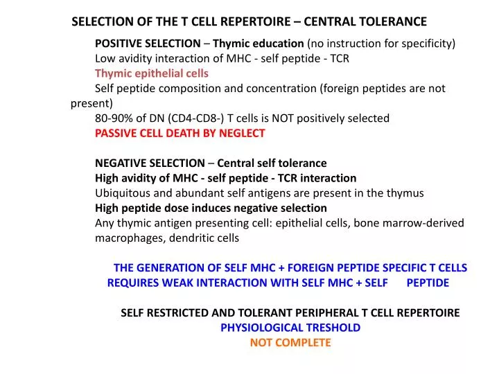
SELECTION OF THE T CELL REPERTOIRE – CENTRAL TOLERANCE POSITIVE SELECTION – Thymiceducation (no instructionforspecificity) Lowavidityinteraction of MHC - selfpeptide - TCR Thymicepithelialcells Selfpeptidecomposition and concentration (foreignpeptidesarenotpresent) 80-90% of DN (CD4-CD8-) T cells is NOT positivelyselected PASSIVE CELL DEATH BY NEGLECT NEGATIVE SELECTION – Centralselftolerance Highavidity of MHC - selfpeptide - TCR interaction Ubiquitous and abundantselfantigensarepresentinthethymus Highpeptidedoseinducesnegativeselection Anythymicantigenpresentingcell: epithelialcells, bonemarrow-derived macrophages, dendriticcells THE GENERATION OF SELF MHC + FOREIGN PEPTIDE SPECIFIC T CELLS REQUIRES WEAK INTERACTION WITH SELF MHC + SELF PEPTIDE SELF RESTRICTED AND TOLERANT PERIPHERAL T CELL REPERTOIRE PHYSIOLOGICAL TRESHOLD NOT COMPLETE
γδT-cells • MHC-independent, CD1c and CD1d dependent • Double megative • comprise up to 50% of the intra-epithelial lymphocyte population • Expanded in intracellularbacterialinfections(Mycobacterium tuberculosis and Listeriamonocytogenes), extracellularinfections (Borreliaburgdorferi) • a population that is expanded in certain disease states such as celiac disease Pierre Vantouroutand Adrian Hayday88 | FEBRUARY 2013 | VOLUME 13
THE ROLE OF PROFESSIONAL ANTIGEN PRESENTING CELLS IN THE IMMUNE RESPONSE Infectiousdiseases Tissuetransplantation Elimination of tumors Autoimmunediseases Gatekeeperfunction Sensingpathogens Primingadaptiveimmuneresponses Maintenance of selftolerancetoselfstructures
Dendriticcellsaresensors, gatekeepers and messengers Activationinducesa phenotypeessentialfor theinitiationof theadaptiveimmuneresponse MHC class II molecules stained green lysosomalprotein stained red
CONTACT OF DENDRITIC CELLS AND T - LYMPHOCYTES IN LYMPHOID ORGANS Activateddendriticcellsactasprofessionalantigenpresentingcells MHC-peptidecomplexes1. signal STRANGER Co-stimulatorymolecule2. signal AMPLIFICATION Cytokines3. signal DANGER Theyareinclosecontactwith specific T lymphocytes
INTERDIGITATING RETICULAR (MATURE DENDRITIC) CELL IN T CELL AREAS OF LYMPH NODES NUCLEUS T CELL T CELL CYTOPLASM
Cell-surface molecules of the immunoglobulin superfamily initiate lymphocyte adhesion to professional antigen-presenting cells B. Transient interactions are stabilized by Ag-binding A. Initial contact A DC-specificintercellularadhesion molecule-3 grabbingnonintegrin (DC-SIGN).
CHANGES OF TISSUE ENVIRONMENT INDUCES THE ACTIVATION OF MACROPHAGES AND DENDRITIC CELLS Phagocytosis and degradation of backteria (LPS, TLR) DANGER SIGNAL Macrophage Activated macrophage Monocyte Dendritic cell Activated dendritic cell Virus, extracellular pathogens, inflammatory cytokines (LPS, TLR) DANGER SIGNAL LYMPHOID TISSUE TISSUE BLOOD
Effector and memory T cells Lymphatics Activated DC Inflammation Pathogen Naive T cells ANTIGEN CIRCULATION Tissue DC ACTIVATION AND MIGRATION OF DENDRITIC CELLS TISSUE LYMPH NODE TISSUE DC AND T CELLS ENCOUNTER T CELL ACTIVATION
Capture of an Ag-Specific T Cell by an Ag-Bearing DC Rapid DC Migration in the Subcapsular Space Bone-marrowderivedDCs (either 5 µM CFSE, green)or (50 µMCellTrackerBlue, blue) wereinjectedintothefootpad of a C57BL/6 mouse, followed 18 hourslaterbyintravenousinjection of freshlyisolatedpolyclonalCD4+ T cells(5 µM SNARF, red) and CD8+ T cells(5 µM CFSE and 5 µM SNARF, yellow). The draining LN wasremoved 6 hoursafterinjection Bone-marrowderivedDCs (yellow) werepulsedwith 1 µMOva 4 peptide and 10 µMOvafor 1 hourat 37oC, theninjectedintothefootpad of a C57BL/6 recipient. Thiswasfollowed 6 hourslaterbyi.v. co-injection of OT-I CD8+ T cells(5 µM CFSE, green) and OT-II CD4+ T cells(5 µM SNARF, red). Huang et al Immunity 2004
Morphology of plasmacytoid dendritic cells IPC/DC2 pDC monocyte In human TLR9 is onlyexpressedin pDCs Scanning EM Transmission EM DC-specificintercellularadhesion molecule-3 grabbingnonintegrin (DC-SIGN).
Migration Pathways of PDC/IPC versus mDC into a lymph node mDC: afferent lymphatics IPC: HEV Both migrateintotheT-cellrichareas PDC efficientlycrosspresentexogenousantigens to CD8+ T-cells
PLASMACYTOID DENDRITIC CELLS AS PROFESSIONAL TYPE I INTERFERON SECRETING CELLS Enhanced NK cell cytotoxic activity TLR4 Vírus infection TRAM TRIF TLR7 TLR8 TLR9 TLR3 TRIF MyD88 TANK IRAK-1 Activation of and γδ T cells TRAF-6 RIG-1 IKKε TBK1 IFN-β IFN-α1 Cross-presentation by conventional dendritic cells is enhanced IRF-3 IRF-5 IRF-7 IRF-7 Ig production by B cells is induced Type I interferon receptor
Plasmacytoid DCs control the function of many immunocytes IFNα is impotant in SLE pathology HIV infects PDC Role in immune response and in the pathogenesis of autoimmune diseases and cancer