Download
1 / 73
740 likes | 992 Views
Management of Postpartum Hemorrhage. Done by Group C2. Presented by :. Dr.Ghassan A. Barayan , MBBS. Case Hx. G5 P4 + 0 lady , in active labour pain . Admitted to the hospital and she delivered a 3.8 kg baby. Immediately after the placental delivery she had heavy vaginal bleeding . DDX

E N D
Management of Postpartum Hemorrhage Done by Group C2 Presented by : Dr.GhassanA. Barayan, MBBS
Case Hx • G5 P4 + 0 lady , in active labour pain . Admitted to the hospital and she delivered a 3.8 kg baby. Immediately after the placental delivery she had heavy vaginal bleeding . • DDX • management
Maternal mortality • Hemorrhage • Infection • Hypertension • Thromboembolism The single most Important cause of Maternal death Every year, 140.000 women die because of postpartum hemorrhage – one every 4 minutes
Definition • Estimated blood loss with vaginal delivery: > 500cc • Estimated blood loss with C/S: > 1000cc • 1ry : within 24h after delivery • 2ndry : after 24h up to 6 - 12 weeks
Avoid underestimating significant bleeding Don’t underestimate EBL Don’t disregard slow but unrelenting trickle If signs of shock* without high EBL, must consider hidden bleeding (internal injury, hidden hematoma) Requires urgent treatment
Epidemiology Incidence : 4 % 88 % of deaths are seen in the first 4 hrs Abdominal or pelvic bleeding can be hidden
Early postpartum hemorrhagecauses Uterine atony Genital tract trauma Retained placental tissue Low placental implantation due to relative musculature in the LUS so insufficient control of bleeding Uterine inversion Coagulation disorders Abruptioplacentae Amniotic fluid embolism Retained dead fetus Inherited coagulopathy
Four “Ts”* Tone . . . . . . - Uterine atony Tissue . . . . . - Retained products Trauma . . . . - Tears, abrasions Thrombin . . . - Clotting disruptions
Clinical presentation • The patient whether presents with Vaginal Bleeding OR Severe hypovolemic shock
“Tone: Think of Uterine Atony” • Uterine atony causes 80% of hemorrhage • failure of uterus to contract after placental separation
Hx : identify risk factor Uterine overdistention: -multiple gestation -polyhydromnios - fetal macrosomia Prolonged oxytocin administration Prolonged labor Precipitous labor (last <3h ) Halogenated anesthesia Magnesium sulfate ttt of pre-eclampsia Chorioamnionitis Uterine fibroids Grand multiparity
Bimanual Uterine Exam Confirms diagnosis of uterine atony: the fundus is soft “ boggy “ , poorly contracting
Initial Assessment (orders) • Remember ABCs • Check the vitals and correct the shock state • Start a large-bore IV • Obtain CBC • Platelet count , PT , PTT • Blood crossmatch (keep 4 units of packed RBCs in hand ) • IV infusion of norm.saline or ringer lactate (it’s best to avoid glucose containing fluids) • 3ml of crystalloid / ml of EBL
Initial Assessment (orders).cont. • Insert urethral catheter to monitor urine output • Blood and blood product transfusion may be required if • blood loss is continuing, • if the blood volume lost is over 30%, • or if the patient’s clinical status reflects developing shock despite aggressive resuscitation
Management Bimanual Uterine massage is often adequate for stimulating uterine involution. medications
Oxytocin ( pitocin ) • promotes rhythmic contractions of the upper uterine segment • Give IM or IU, no rapid IV infusion (Can cause BP) • 10 – 40 U in 1 L NS at 250cc/h. / IV • Can get Antidiuretic effect in very high doses
Methylergonovine • Methergine 0.2 mg IM only • Max. of 3 doses • contracts both upper and lower uterine segments tetanically • causes vasoconstriction and hypertension • contraindicated in hypertension • side effects: HTN, nausea, vomitting
Prostaglandins F2 • Hemabate 0.25mg intramyometrial q 15min • Rapid response 3-10 minutes • Max. 8 doses • Avoid in asthmatic patients. • This is 80% to 90% effective in stopping PPH in cases that are refractory to oxytocin and ergometrine
misoprostol Cytotec Recent data indicates that it can be used as 1st line 800 – 1000 mcg rectally
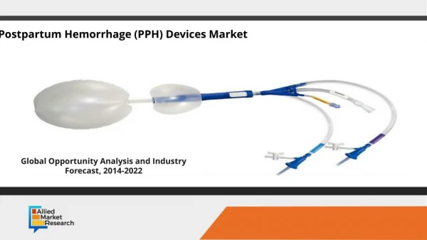
Other measures • Uterine packing or tamponade • When uterotonic agents fail • Useful in cases of placenta praevia or accreta • Either gauze or Foley, Sangestaikinblakemore tube • Recent reports of large series have confirmed the high success rates of balloondevices • Uterine artery embolizationby placing angiocatheter and injecting thrombogenic material
Exploratory laparotomy : • B-lynch technique : effective in uterine atony • Bilateral uterine artery ligation • Hypogastric artery ligation • Suprecervical or TAH is the definitive treatment for intractable PPH
Human recombinant factor VIIa • New treatment to control severe , life threatening hemorrhage • It acts on the extrinsic clotting pathway • Cessation of bleeding occurs in 10-40 minutes
2-Tissue: Retained placenta • Prolong 3rd stage of delivery( > 30 minutes) seen in ~ 6% of deliveries. • Prior retained placenta. • Prior manual removal of placenta • Prior C/S, curettage p-pregnancy, uterine infection, and increased parity. • Prior C/S scar & previa (25%) • Most patients have no risk factors.
US evaluation of retained tissue should be performed before uterine instrumentation
Brandt-Andrews maneuver To determine if the placenta has separated Firm traction is applied to the umbilical cord with one hand while the other applies suprapubiccounterpressure
Abnormal adherent placenta Caused by missing or defective decidual layer . • Placenta Accreta: Placenta adherent to myometrium. • Placenta Increta: myometrial invasion. • Placenta Percreta: penetration of myometrium to or beyond serosa.
Placenta Accreta: • Most common type. • With incidence of 15-25 % if previous C/S or previa • The incidenceincrease as the no. of previous C/S increased • Prior uterine surgery and placenta praevia in the current pregnancy r 2 important RF – • US and color doppler • TAH is the definitive treatment
Placenta accreta and uterine atony are the 2 commonest causes of postpartum hysterectomy
Removal of Abnormal Placenta • Oxytocin 10U in 20cc of NS placed in clamped umbilical vein. • If this fails, get OB assistance. • Check Hct, type & cross 2-4 u. • Two large bore IVs. • Anesthesia support.
Removal of Abnormal Placenta • Relax uterus with halothane general anesthetic and subcutaneous terbutaline. • Bleeding will increase dramatically. • With fingertips, identify cleavage plane between placenta and uterus. • Keep placenta intact. • Remove all of the placenta.
Removal of Abnormal Placenta • If successful, reverse uterine atony with oxytocin, Methergine, Hemabate. • Consider surgical set-up prior to separation. • If manual removal not successful, large blunt curettage or suction catheter, with high risk of perforation, so to be done on US guidance • Consider prophylactic antibiotics.
Trauma (3rd “T”) (cervical, vaginal and preineal laceration) Suspected if bleeding persists and uterine fundus is contracting. Episiotomy Hematoma Uterine inversion Uterine rupture In C/S LT laterl extension of the incision can damage ascending branches of the uterine a while inferior extension damages the cervical branches.
Genital tract Trauma Risk factors include: Instrumented deliveries forceps or vaccum. Primiparity. Pre-eclampsia. Multiple gestation, fetal macrosomia. Prolonged second stage, precipitous delivery VBAC Clotting abnormalities.
Repair of cervical laceration At the two lateral angles while dilating the first stage. Repair lacerations quickly. Place initial suture above the apex of laceration to control retracted arteries.
Vulvar Hematomas • more likely w/ operative vaginal deliveries and episiotomies • excessive unilateral swelling and pain • Hematomas less than 3cm in diameter can be observed expectantly. • If larger or expanding , incision and evacuation of clot is necessary. • Irrigate and ligate bleeding vessels. • With diffuse oozing, perform layered closure to eliminate dead space. • Broad spectrum antibioticsshould be given
Limited from spread by fascia lata • Central tendon of the perineum prevents it from crossing the midline
Retroperitoneal heamtoma • more likely after C/S delivery – symptom may be shock, excess flank pain.
Uterine Inversion • Rare: ~1/2500 deliveries • Turning inside out of the uterus • C/F: • Acute abdominal pain • Blue-gray mass protruding from vagina. • Suspect if shock disproportionate to blood loss