Download
1 / 30
300 likes | 481 Views
** Digestive System ** Small/Large Intestine Liver A&P Lecture Notes Pages 98-103. Liver, Bile ducts, Pancreas and Small Intestine. Figures from: Marieb, Human Anatomy &Physiology, Pearson, 2013. Three Parts of Small Intestine. Figure from: Hole’s Human A&P, 12 th edition, 2010.

E N D
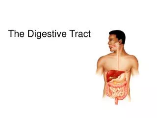
** Digestive System ** Small/Large IntestineLiver A&P Lecture Notes Pages 98-103
Liver, Bile ducts, Pancreas and Small Intestine Figures from: Marieb, Human Anatomy &Physiology, Pearson, 2013
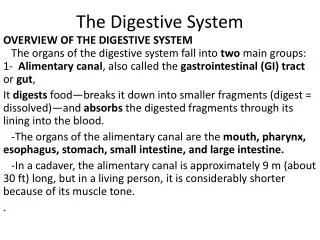
Three Parts of Small Intestine Figure from: Hole’s Human A&P, 12th edition, 2010 “Mixing bowl”; acid neutralization Bulk of chemical digestion and nutrient absorption occurs here The ‘bowel’ consists of the small and large intestines. Vitamin B12 absorption Main functions of small intestine: 1) chemical digestion 2) absorption of nutrients (90%) from chyme
Blood Supply and Drainage of Small Intestine Figure from: Martini, Anatomy & Physiology, Prentice Hall, 2001
Wall of Small Intestine Figure from: Hole’s Human A&P, 12th edition, 2010 Plicae circulares – permanent circular folds of mucosa that further increase surface area for absorption – do not flatten out with distention like rugae of stomach. Especially prominent in lower duodenum and upper jejunum Submucosa of duodenum contains mucus-secreting glands (Brunner’s glands) that protect the small intestine
Intestinal Villi & Glands Enterocyte = Intestinal Cell Figure from: Saladin, Anatomy & Physiology, McGraw Hill, 2007 Intestinal glands secrete an abundant watery fluid that helps absorb products of digestion. They also contain enteroendocrine cells (enterokinase, gastrin, secretin, CCK)
Intestinal Epithelium Figure from: Hole’s Human A&P, 12th edition, 2010 Microvilli further increase the surface area available for absorption in the small intestine Form a ‘brush border’ on the intestine Digestive enzymes are embedded in the membrane of microvilli Main function of plicae, villi, and microvilli is to increase the surface area for absorption (from about 3.6 ft2 to about 2200 ft2!)
Secretions of Small Intestine • peptidase – breaks down peptides into amino acids • sucrase, maltase, lactase – break down disaccharides into monosaccharides • intestinal lipase – breaks down fats into fatty acids and glycerol • enterokinase – converts trypsinogen to trypsin • gastrin/somatostatin – hormones that stimulate/inhibit acid secretion by stomach • cholecystokinin (CCK) – hormone that inhibits gastric glands, stimulates pancreas to release enzymes in pancreatic juice, stimulates gallbladder to release bile, and relaxes hepatopancreatic sphincter (of Oddi) • secretin – stimulates pancreas to release bicarbonate ions in pancreatic juice; stimulates gall bladder to release bicarbonate-rich bile Brush border See Table 23.32 in Marieb for a great summary of digestive enzymes
Movements of the Small Intestine Movements in local segments can occur without stimulation by parasympathetic NS. However, nervous stimulation accelerates segmentation and peristalsis. • peristalsis – pushing movements • segmentation – ringlike contractions that aid in mixing and slowing peristalsis • overdistended or irritated wall triggers “peristaltic rush” resulting in diarrhea “Long distance” movements are triggered by stomach filling: - gastroenteric reflex (↑ motility and secretion along length of small intestine) - gastroileal reflex (relaxation of ileocecal sphincter)
Absorption in the Small Intestine • monosaccharides and amino acids • through facilitated diffusion and active transport • absorbed into blood • electrolytes and water • through diffusion, osmosis, and active transport • absorbed into blood • vitamins • fat-soluble dissolve in dietary fats (vit A,D,E,K) • Water-soluble through diffusion, except B12 (active transport) • Vitamin K (large intestine) – with other lipids • absorbed into blood
Absorption of Fats in the Small Intestine Figure from: Hole’s Human A&P, 12th edition, 2010 • fatty acids and glycerol • several steps • absorbed into lymph into blood Chylomicrons contain TG, cholesterol, and phospholipids
Large Intestine Figure from: Martini, Anatomy & Physiology, Prentice Hall, 2001 *
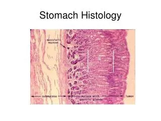
Histology of the Large Intestine Figures from: Hole’s Human A&P, 12th edition, 2010 Walls of large intestine are much thinner than the small intestine, however, the lumen is larger Note lack of villi and presence of numerous goblet cells (mucus) No enzymes produced; any digestion is from previously introduced enzymes or bacteria
Functions of Large Intestine • little or no digestive function • absorbs water, bile salts, and electrolytes • secretes mucus (lubrication, binding, protection, pH) • conversion of bilirubin (uro- and stercobilinogen) • houses intestinal flora (~800 species of bacteria) and absorbs vitamins liberated by bacterial action (K, B5, and Biotin); produces intestinal gas (flatus) • forms and stores feces • carries out defecation
The Rectum, Anal Canal, and Anus Figure from: Hole’s Human A&P, 12th edition, 2010 Temporary storage of fecal material in rectum triggers the urge to defecate Internal anal sphincter is usually contracted but relaxes in response to distension. External sphincter must be tensed reflexively to retain feces Rectal valves Procto- = anus or rectum (Keratinzed strat. squamous epithelium)
Movements of Large Intestine • slower and less frequent than those of small intestine • mixing movements (haustral churning every 30 min) • mass movements - usually follow meals (stimulated by distension of stomach and duodenum) • gastrocolic reflex • duodenocolicreflex • peristaltic wave from transverse colon through rest of large intestine
Parasympathetic Defecation Reflex Figure from: Saladin, Anatomy & Physiology, McGraw Hill, 2007 Note that this reflex opens the internal sphincter and closes the external sphincter Need voluntary relaxation of the external sphincter for defecation
Feces • water (75%), solids (25%) • electrolytes • mucus • bacteria (30% of solids) and sloughed epithelial cells • bile pigments altered by bacteria provide color (mainly urobilins and stercobilins) • odor produced by bacterial compounds (indoles and skatoles, phenols, H2S, ammonia) • indigestible materials
Major Organs of Digestive System Figure from: Saladin, Anatomy & Physiology, McGraw Hill, 2007 • Organs can be divided into the: • Digestive tract (primary) (alimentary canal); tube extending from mouth to anus (about 30 ft.) • Accessory organs; teeth, tongue, salivary glands, liver, gallbladder, and pancreas
Liver [ Hepat(o)- ] Round ligament is part of the falciform ligament that divides the lobes; remnant of fetal umbilical vein. Note that the vena cava does not enter the liver; it passes by Figure from: Martini, Anatomy & Physiology, Pearson Education, 2004
Arterial Supply and Venous Drainage of Liver Figure from: Martini, Anatomy & Physiology, Prentice Hall, 2001
Hepatic Lobule Hepatic lobules are the functional units of the liver (>100,000) Figure from: Saladin, Anatomy & Physiology, McGraw Hill, 2007
Paths of Blood and Bile in Hepatic Lobule Figure from: Hole’s Human A&P, 12th edition, 2010 Liver’s role as an accessory organ in digestion is production of bile Sinusoid Hepatic portal vein → sinusoids → central vein → hepatic veins → inferior vena cava Hepatic artery
Liver Functions (over 200!) • Three general categories of function 1) Metabolic regulation • Interconversion of carbohydrates, lipids, amino acids • Removal of wastes • Vitamin and mineral metabolism • Drug inactivation • Storage of fats, glycogen, iron, vit A/B12/D/E/K 2) Hematological regulation • Phagocytosis and antigen presentation; ab removal • Synthesis of plasma proteins • Removal of circulating hormones • Removal of worn-out RBCs (Kupffer cells) • Removal or storage of toxins 3) Synthesis and secretion of bile (role in digestion) Know items in red
Composition of Bile (Chole-) Yellowish-green liquid continually secreted by hepatocytes • water • bile salts (bile acids) • derived from cholesterol • emulsification of fats (increases surface area for digestive enzymes) • helps absorption of fatty acids, cholesterol, and fat-soluble vitamins • 80% are recycled (reabsorbed and reused) – enterohepatic circulation of bile • 20% excreted in feces (disposes of excess cholesterol) • bile pigments (bilirubin and biliverdin from breakdown of RBCs) • electrolytes
Gallbladder [Cyst(o)-] Figure from: Martini, Anatomy & Physiology, Prentice Hall, 2001 Main function is to store and concentrate bile between meals, and release bile under the influence of CCK
Regulation of Bile Release from GB Figure from: Hole’s Human A&P, 12th edition, 2010 • fatty chyme entering duodenum stimulates the GB to release bile (via CCK) Secretin causes the bile ducts (and pancreatic ducts) to secrete bile rich in HCO3-
Actions of Cholecystokinin (CCK) on Digestion Figure adapted from: Barrett, K., Gastrointestinal Physiology, Lange, 2006 CCK Contraction of Gallbladder Secretion of pancreatic enzymes Reduced emptying of stomach Relaxation of hepatopancreatic sphincter Protein, CHO, lipid absorption and digestion Matching of nutrient delivery to digestive and absorptive capability
Pancreatic Juice • pancreatic amylase – splits glycogen into disaccharides • pancreatic lipases – break down triglycerides • pancreaticnucleases – digest nucleic acids • bicarbonate ions – make pancreatic juice alkaline (pH = 8) and neutralize acid coming from stomach • Pancreatic proteolytic enzymes…