Download
1 / 52
570 likes | 847 Views
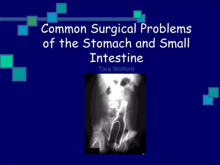
Common Surgical Problems of the Stomach and Small Intestine Tara Wofford. Links to Helpful Online Tutorials. An Approach to Abdominal Plain Films: http://www.learningradiology.com/lectures/gilectures/plainabdomenflashpage.htm

E N D
Common Surgical Problems of the Stomach and Small IntestineTara Wofford
Links to Helpful Online Tutorials • An Approach to Abdominal Plain Films: http://www.learningradiology.com/lectures/gilectures/plainabdomenflashpage.htm • Abnormal Bowel Gas Patterns: http://www.learningradiology.com/notes/ginotes/pictorialbowelgas.htm • Recognizing SBO, LBO, and Paralytic Ileus: http://www.learningradiology.com/medstudents/recognizingseries/recogobstructflashpage.htm
Objectives • To become familiar with abdominal anatomy on a radiograph and CT image. • To be able to identify the radiologic signs of pathology of the stomach and small intestine. • To be able to evaluate the presence of common surgical problems in these organs.
Anatomy of the Stomach • The stomach has four parts (cardia, fundus, body, and pylorus) and two crurvatures. • The gastric mucosa forms longitudinal folds called rugae. • The stomach is bordered anteriorly by the diaphram, left lobe of the liver and ant. abd wall. Posteriorly it is bordered by the omental bursa and pancreas
Barium X-Rays • A barium swallow, also called an upper GI series, is an examination of the esophagus and stomach using barium to coat the walls of the upper digestive tract so that it may be examined under x-ray. • Barium swallows are used to identify any abnormalities such as tumors, ulcers, hernias, pouches, strictures, and swallowing difficulties.
High-Resolution CT scan of stomach • Optimal luminal distension with barium, water, or gas is key for evaluating the gastric wall. • Water-soluble oral contrast is used when perforation is suspected.
Common Surgical Pathology of the Stomach • Peptic Ulcers • Tumors - Adenocarcinoma, Lymphoma • Hiatal Hernia
Peptic Ulcers • Causes: H. Pylori infection, hyperparathyroidism, steroid tx, uremia, stress, burns (curling), cerebral disease (cushing). • Duodenal ulcers are also present in 5-42% of cases. The ratio of DU:GU is 3:1. • Most common location of benign ulcers is the lesser curvature area of body and antrum. • Almost all lesions < 1cm are benign.
Radiologic Findings • Ulcer crater: barium collection on dependent surface penetrates beyond anticipated wall. • Hampton’s line: 1mm thin straight line at neck of ulcer, represents undermined mucosa. • Ulcer collar: smooth, thick, lucent band at neck of ulcer, represents thicked wall. • Ulcer mound: tissue mass surrounding ulcer.
Adenocarcinoma of the Stomach • 24,000 new cases diagnosed each year. • M:F ratio is 2:1 • Risk factors: H. Pylori infection, adenomatous polyps, chronic atrophic gastritis, pernicious anemia and partial gastrectomy.
Radiologic Findings • Conventional CT is not sensitive in early phases compared to HRCT. • Early cases may appear as focal wall thickening with mucosal enhancement during the early arteriovenous contrast phase. • Advanced cases appear as thickened, abnormally enhancing gastric wall, in localized or circumferential pattern,or as a polypoid mass. Ulceration may be apparent as well.
Gastric Lymphoma • The stomach is the most common site for GI lymphoma and is more commonly part of a generalized disease. • 80% of cases are Non-Hodgkins. • Perforation is a major complication occuring in 9-47% of patients
Radiologic Findings which differentiate Lymphoma from Adenoarcinoma • Gastric wall thickness is much greater in lymphoma, with a mean of 4cm. • Adenopathy is more pronounced and lymph nodes larger. • Mural thickening is more homogenous.
Hiatal Hernia • Caused by a weakness or tear in the phrenoesophageal membrane. There are two types: • Sliding hernia, in which the gastroesophageal junction is displaced above the diaphram (includes 99% of cases). • Paraesophageal hernia, in which there is stomach herniating into chest but the GE junction is not effected.
Radiologic Findings • Extension of multiple gastric folds above the diaphram. • Bulbous area of distal esophagus containing contrast. • “Schatzki’s Ring” - a filling defect that marks the position of esophagogastric junction and defines the presence of sliding hernia
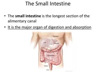
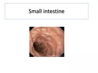
Anatomy of Small Intestine • Includes duodenum, jejunum, and ileum. • The mucosal wall is characterized by circular folds (plicae circulares). • The duodenum has a c-shaped course around the pancreas and is partially retroperitoneal. • Most of the jejunum lies in the LUQ and ileum mostly in RLQ.
Assessing the Abdominal Radiograph • Gas pattern: stomach- always a small amount, small int. will have 2-3 gas filled loops (< 2.5cm in diameter), and rectum- usually has a small amount. • Air-fluid levels: stomach- always present, small int. may have 2-3 levels, usually never in rectum. • Also look for soft-tissue masses and calcifications.
Small vs Large Intestine • Small intestine: located centrally, has circular folds that extend across the lumen, and has a maximum diameter of 2-2.5 cm. • Large Intestine: located peripherally and has haustral markings that do not cross the lumen
Abdominal Anatomy on CT Identify Stomach, small and large intestine, liver, spleen, and descending aorta.
Vascular Supply to the Small Intestine • The duodenum is supplied by both the celiac trunk (proximal to bile duct) and the SMA (distal to the entry of the bile duct). • The jejunum and ileum are supplied by the SMA via 15-18 branches which form arterial arcades that give rise to the vasa recta.
Common Surgical Problems of the Small Intestine • Small Bowel Obstruction • Crohn’s Disease • Acute Mesenteric Ischemia • Intussusception • Gall-Stone Ileus • Tumors - Adenocarcinoma, Lymphoma • Pneumoperitoneum • Superior Mesenteric Artery Syndrome
Small Bowel Obstruction • Pathophysiology: bowel proximal to obstruction dilates with swallowed air and secretions and there is hyperparastalsis. Ischemia can occur from vascular compromise of effected loops. • Most common etiologies: adhesions from prior surgery, hernia, intussesception, gallstone ileus, volvulus, and tumors. • Clinical symptoms: abdominal pain, distention, N/V/D, hyperactive bowel sounds. • Surgery is indicated with s/s of ischemia, peritonitis, or when refractory to conservative tx.
Radiographic Findings • Proximal loops dilated >2.5-3cm • Multiple air-fluid levels • Absence or small amount of gas in colon.
Associated CT Findings • Dilated, fluid filled loops of small bowel proximal to obstruction and collapse of distal bowel. • Signs of ischemia include thickening of bowel wall, stranding of adjacent to small bowel mesesentary or pneumatosis intestinalis.
Crohn’s Disease • Characterized by non-caseating granulomas with transmural inflammation, which can effect any part of the GI tract. • Clinically patients frequently have recurrent diarrhea, occult blood loss, anemia, abd pain, and low-grade fever. • Small intestine is involved 80% of the time, particularly the terminal ileum.
Radiographic findings • Skip lesions - separated by normal areas of bowel. • Squaring of folds, indicating lymphedema. • Apthous ulcers - small nodular filling defects which appear as a mound of edema with central ulceration. • String-sign - marked narrowing of terminal ileum usually from edema, spasm and fibrosis. Proximal dilatation is common.
Associated CT Findings • Bowel wall thickening with skip lesions. • Proliferation of mesenteric fat and lymphadenopathy. • Inflammatory stranding.
Ddx in Crohn’s Dz • Ulcerative Colitis - the entire colon is frequently involved with the terminal ileum spared. • Diverticulitis - diverticula are present, mucosa is intact, and terminial ileum less involved. • TB - cecum is more effected than terminal ilem. • Lymphoma - tumor masses are visualized.
Acute Mesenteric Ischemia • Defined as interruption of blood supply to small or large intestine.(Associated with 70-90% mortality overall) • Causes: embolism (SMA most common), arterial thrombus, venous thrombus, and diffuse mesenteric vasoconstriction due to low cardiac output. • Common clinical symptoms: severe abd pain out of proportion to exam, usually poorly localized, N/V/D, and GI bleeding. • Surgical options: thrombectomy/ embolectomy, arterial bypass, and resection of necrotic bowel.
Radiographic Findings • X-ray is abnormal in 20-60%. • Thumbprinting - (nonspecific) indicates wall edema and hemhorrage in the this setting. • Pneumatosis, portal vein gas, pneumoperitoneum - indicates infarcted bowel. Pneumoperitoneum Pneumatosis
Possible CT Findings • Bowel wall thickening indicating edema or hemorrhage. • Lack of enhancement in wall indicated infarction. • Pneumatosis, portal vein gas, pneumoperitoneum. • Intraluminal thrombus in involved vessel.
Intussusception • Most common cause of bowel obstruction in kids but much less common in adults. • In adults there is usually an associated cause such as a mass, polyp, or adhesions. • There are three types: enteroenteric, ileocolic, and colocolic. • CT characteristics include a target-shaped mass enveloped with a thick outter rim of soft tissue representing edematous bowel wall.
Gallstone Ileus • Occurs when a gallstone erodes into GI tract and causes obstruction. • Dilated loops of small intestine are seen, with air in the biliary tree and gallbladder. • The stone is usually located in the terminal ileum but can be anywhere along small intestine.
Adenocarcinoma of the Small Intestine • Most often this lesion arises in the proximal jejunum. • Risk factors: hx of Crohn’s, sprue, Peutz-Jeghers, and duodenal/jejunal bypass surg, among others. • Common types are infiltrative (bowel obstruction) and ulcerative (bleeding). • On CT the tumor appears as eccentric focal mass or circumferential bowel wall thickening in a short segment.
Lymphoma of the Small Intestine • Occurs most often in the ileum where there is more lymph tissue. • Risk factors: immunocompromised or suppressed state, celiac sprue, and CLL. • Small intestine is the second most common site in the GI tract for lymphoma.
CT Findings • The typical patterns are aneurysmal, constrictive, nodular, and ulcerative. • There is frequently asymmetric wall thickening (>2cm), aneurysmal dilatation, polyploidal mass, abdominal lymphadenopathy. • Tissue density in the thickened bowel is relatively homogenous.
Pneumoperitoneum • Defined as free air in abdominal cavity. • Causes: • disruption of a hollow viscus from trauma, iatrogenic perforation, or GI tract disease. • Extension from chest. • Via female GU tract. • Through peritoneal surface via a procedure. • Intraperitoneal source, such as abscess rupture or gas-forming microbes.
Key Radiologic findings • “Rigler’s sign” or double-wall sign, which appears as air on both sides of bowel wall (usually indicating > 1000 ml of free air). • RUQ is the best place to look for small air collections. These appear as lucency over liver. • With a larger gas collection the patient may have abdominal distension and lack a gastric air-fluid level.
Superior Mesenteric Artery Syndrome • Compression of the third (transverse) portion of duodenum against the aorta by the SMA. • This results in chronic or intermittent acute complete or partial obstruction.
42 y.o. woman presents with abd pain, distension, and nausea. What is the most likely problem? A. Mesenteric Ischemia B. Small Bowel Lymphoma C. Small Bowel Obstruction
42 y.o. woman presents with abd pain, distension, and nausea. What is the most likely problem? C. Small Bowel Obstruction
65 y.o. man c/o rapid onset of diffuse abd pain combined with vomiting, diarrhea, and blood in the stool. What is the likely problem? A. Perforated Gastric Ulcer B. Acute Mesenteric Ischemia C. Small Bowel Obstruction.
65 y.o. man c/o rapid onset of diffuse abd pain combined with vomiting, diarrhea, and blood in the stool. What is the likely problem? B. Acute Mesenteric Ischemia
32 y.o. woman with recurrent moderate epigastric pain, hematemesis, and anorexia. What is the likely problem? A. Gastric Ulcer B. Gastric Adenocarcinoma C. Gastric Lymphoma
32 y.o. woman with recurrent moderate epigastric pain, hematemesis, and anorexia. What is the likely problem? A. Gastric Ulcer
42 y.o. man with recurrent crampy abd pain combined with weight loss, diarrhea, and fever. What is the likely cause? A. Small Bowel obstruction B. Acute Mesenteric Ischemia C. Crohn’s disease