Download
1 / 36
360 likes | 456 Views
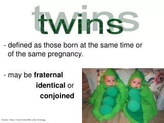
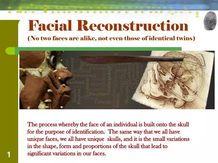
Facial Reconstruction (No two faces are alike, not even those of identical twins).

E N D
Facial Reconstruction (No two faces are alike, not even those of identical twins) The process whereby the face of an individual is built onto the skull for the purpose of identification. The same way that we all have unique faces, we all have unique skulls, and it is the small variations in the shape, form and proportions of the skull that lead to significant variations in our faces.
Facial Reconstruction In this section of Forensics, you will: • Learn the anatomy of the head • Work the Manchester Method of forensic reconstruction • Understand the anatomical roots of facial expression
History of Facial Reconstruction • 7000 BC in Neolithic Jericho – Skulls of the deceased were plastered and adorned with cowrie shells mimicking living tissue. These were used for spiritual purposes rather than for identification. These types of skulls have also been found in Meso-America and New Guinea. • Aristotle (384-322 BC) – first to apply physiognomy (the deduction of • character from facial morphology) in his book Historiaanimalium • Bertillon (1877) – Paris Police force. He kept face and body • measurements on all criminals.
Facial Reconstruction - History • 1907 – 1970 Mikhail Gerasimov spent his young years collecting and bones and reconstructing. He worked mainly with the cranium working with his knowledge of the individual traits. Some of his work is still used today. • Gatliff and Snow (1970) – developed the “American Method” of facial reconstruction based only on tissue depth. • Enlow (1982) – Facial differences and additional details such as eye color, skin color, hair color and texture can suggest the sex, ethnic origin and age of a face. • Richard Neave (1997) – developed the “Manchester Method” based on the relationship the soft and hard tissues of the face.
Methods of Facial Reconstruction • American Method – developed by Betty Gatliff and Clyde Snow. It relies primarily on tissue depth data and does not require the application of fleshy tissues such as muscle. • Manchester Method – developed by John Prag and • Richard Neave. This method combines tissue depth, • anatomy, and careful observations of the skull. This • method was developed at the University of Manchester • due to the work done on Egyptian mummies. • We will be using this method!
Navigating the Cranium - Bones The skull is made up of 22 bones consisting of 14 facial bones and 8 cranial bones. These are the required bones! Calvarium Frontal Eminence Infraorbital Margin Supraorbital Ridge Mandible Mastoid Process Maxilla Parietal Eminence Temporal Bone Zygomatic Bone
Navigating the Cranium - Bones Calvarium Infraorbital Margin Frontal Eminence Supraorbital Ridge
Navigating the Cranium - Bones Mastoid Process Parietal Eminence Mandible Maxilla
Navigating the Cranium - Bones Temporal Bone Zygomatic Bone
Navigating the Cranium - Muscles • Frontallis • Septum Cartilage • Procerus • Orbicularis oculi • Orbicularis oris • Zygomatic major • Zygomatic minor • Mentalis • Masseter • Temperal
Navigating the Cranium - Muscles Orbicularis oculi Frontalis Orbicularis oris Septum Cartilage Zygomatic minor Procerus Zygomatic major
Navigating the Cranium - Muscles Depressor Anguli oris Depressor Anguli inferior Mentalis Masseter Temporal
Navigating the Cranium – Glands Parotid glands – salivary gland Cheeks are irregular in shape and glands fill them in. EX. Squeeze your cheeks near your masseter with your hand and pull forward. You can feel the increase in saliva in your mouth.
Navigating the Cranium - Adipose • Adipose tissue • fatty tissue, • determined by genetics. • influenced by race, gender • changes with age particularly along the jawline. • correlates to body weight. Weight can be determined by clothes left on the body • cultural background. Tissue depth markers indicate body fat on an average. *not on nose or zygomatic bone *lateral sausage shape near the supraorbital ridge present on women.
Assess the gender, age and race To reconstruct, one must assess the skull for an accuracy of gender, age estimated to within five years and one of three racial groups (Caucasian, Negroid and Mongoloid) Gender MALE FEMALE • Skull • Narrow • Skull • Rounder • Prominent Occipital Bone • Forehead • Higher, smoother, vertical, more round • Forehead • Less bossed • Supraorbital Ridges • Strongly developed • Orbital Margins • round • Glabella (brow) • Larger • Nasal aperture • Narrow • Orbital Margins • Rectangular • Cheekbones • Heavier -Ushaped • Cheekbones • parabolic Mandible - thicker
Assess the gender, age and race Age 1. Bones begin to fuse at approximately 16 years. Sacrum (base of spine, consists of 5 vertebra) – fuse at age 16 -23. Sutures - plates are not sutured in young children. Soft areas are apparent in children 0-3 yrs of age. Fusion of sutures usually begin at age 17 and complete at 25 years. Old age (65 and older) sutures are almost hidden. Basilar suture – (base of skull) the best area for age determination. 18 – 24 yrs
Assess the gender, age and race Age 2. Teeth are the primary assessment of age of skull • Juvenile • Central incisor 6-9 months • Lateral incisor 7-11 months • Canine 16-20 months • First molar 10-16 months • Second molar 20-26 months • Permanent Adult • Central incisor 6-8 years • Lateral incisor 7-9 years • Canine 9-17 years • First premolar 10-12 years • Second premolar 11-13 years • First molar 6-7 years • Second molar 11-13 years • Third molar 17-25 years Teeth – (dentition) wear and tear is relatively precise although eating and lifestyle is an influence. Smoking will damage teeth and consistent use of a pipe can cause breakage.
Assess the gender, age and race Race • Most commonly used racial groups: • Caucasoid – Europeans • Negroid – West and South Africa • Mongoloid – Asiatics, Inuit (Canadian, • Alaskan) and Native Americans
Assess the gender, age and race Race Caucasoid • Long narrow shape • Narrow nasal aperture • Depressed glabella • Sharp cheekbones • Prominent frontal aspect • Prominent chin • Steeper forehead • Lower face
Assess the gender, age and race Race Negroid • Long head shape • Wide nasal aperture • Sharp upper orbital margins • Low round nasal root • Rounded glabella • Wide interorbital distance • More rounded forehead • Bimaxillary protrusion
Assess the gender, age and race Race Mongoloid • Round head shape • Medium nasal aperture • Rounded orbital margins • Massive cheek bones • Prominent zygomatic bones • Straight nasal profile • Flatter face • Shallow orbitals • Upright forehead • Less protruding nose • Narrowest mouth • Smallest upper lip
So……What do we have??? AGE?? 40-60 years of age GENDER?? Female RACE?? Caucasion
Let’s Begin!Tissue Thickness in mm (Stephen/Simpson) • Locate these on your skull. • Cut eraser to the correct mm. • Sand paper area. • Hot glue • Opisthocranion 5.0 • Vertex 3.5 • Grabella 4.75 • Nasion 5.5 • Mid-nasion 3.0 • Rhinion (end of nasais) 2.75 • Sub-nasale 11.00 • Mid-phitrum 8.5 • Labralesuperius(upper lip margin) 9.0 • Labraleinferius (lower lip margin) 10.0 • Mentolabial sulcus (chin-lip fold) 9.5 • Pogonion (mental eminence ) 10.0 • Gnathion 6.0 • Menton (beneath chin) 5.75 • Mid-supraorbital 7.0 • Mid-infraorbital6.0 • Zygomatic arch 7.5 • Gonion 12.00 • Supra canine 9.5 • Infra-canine 10.5 • Supra M 19.25 • Mid-ramus 16.0 • Mid-mandibular border 9.5 • Infra-M 15.5 • Alare curvature point 8.5
Let’s Begin – Tissue Markers • op Opisthocranion 6.0 • v Vertex 4.5 • g Grabella 4.75 • n Nasion 5.5 • mn Mid-nasion 4.0 • rhiRhinion (end of nasais) 2.75 • sn Sub-nasale 13.0 • mp Mid-phitrum 8.5 • lsLabralesuperius(upper lip margin) 9.0 • li Labraleinferius (lower lip margin) 10.0 • mlsMentolabial sulcus (chin-lip fold) 9.5 • pgPogonion (mental eminence 10.0 • gnGnathion 8.5 • m Menton (beneath chin) 5.75 • mso Mid-supraorbital 7.0 • mio Mid-infraorbital6.0 • acpAlare curvature point 9.5 • go Gonion 12.0 • z Zygomatic arch 7.5 • sC Supra canine 9.5 • iC Infra-canine 10.5 • sM2 Supra M 19.25 • mr Mid-ramus 17.5 • mmb Mid-mandibular border 10.5 • iM2 Infra-M 15.5 v v mso mso g n g mn mso op n rhi mio mn mio rhi z z mso acp z acp sn acp mp sM2 sM2 sn mr sM2 mr mp sC mr ls sC ls sC li iM2 iM2 iC iC go go go li iC iM2 mls mls pg mmb pg mmb gn mmb gn m m
Let’s Begin – Muscle Insertion http://www.youtube.com/watch?v=6G0LvImAGAg Order of Deposition: • Tissue Markers • Eyes • Muscles • Nose • Lips • Skin • Ears
Eyes Place clay in the back of the socket Locate the lacrimal crest (raised bony ridge where the tear duct enters the orbit) Locate the malar tubercle ( bump in the contour, 9mm below the zygomatic suture)
Eyes The eyeball should be positioned so that the flat plane of the iris is touching a tangent taken from the mid- supraorbital point to the mid-infraorbital point.
Nose 1. Project a medial line downward from downward “curve” of the nasal bone. 2. Project a line from the nasal spine outward. 3. Connect the lines (popsicle sticks) with hot glue.
Mouth – lip thickness Measure the length of the enameled are on the teeth • White Europeans Upper lip = .4 + .6 X (upper teeth height) Lower lip = 5.5 + .4 X (lower teeth height) • Asians Upper lip = 3.4 + .4 X (upper teeth height) Lower lip = 6 + .5 X (lower teeth height)
Mouth • Place the chelion points (corners of the mouth) at the junction of the top canine and the 1st premolar. • Place them about half way down the enameled surface of the teeth. 1st Incisor 2nd Incisor 1st Premolar canine
Ears • Small mastoid process, which are directed inside the skull, suggest small ears that are close to the head. • Massive prominent mastoids indicate large spread-out ears. • Mastoid process are directed downward = attached lobe. • Mastoid process point forward = lobe is free
Glands and Adipose Tissue Fill in adipose tissue under risorius muscle and zygomatic major and zygomatic minor. Fill in the parotid gland Fill in adipose where necessary.
Skin 1. Roll clay into sheet about 3mm thick. (Mist clay if it sticks to the roller) 2. Keep skin and eyes extremely thin. • Find individuals online of a variety of individuals of the same age, race and gender that you estimate of your reconstruction. Use for creases, edges of lips, bags and folds around eyes. DO NOT MUSH. Only push to find the markers!
Eyebrows, hair and attire. • Eyebrows and hair can be added using actual hair, false hair, or by simply molding a texture with a tool. • Attire may be from clothing replicated from the crime scene.