Download
1 / 9
200 likes | 1.07k Views
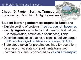
Protein sorting and the Golgi apparatus. The Golgi Apparatus. Because of its large and regular structure, the Golgi apparatus was one of the first organelles described by early light microscopists.

E N D
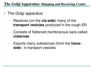
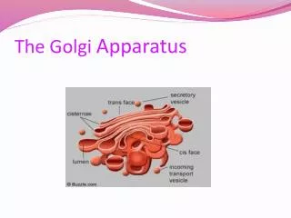
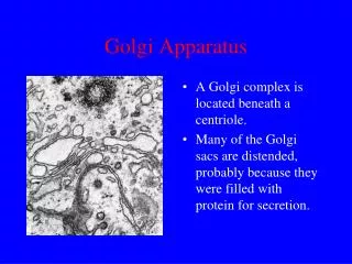
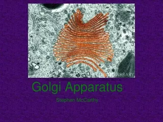
The Golgi Apparatus • Because of its large and regular structure, the Golgi apparatus was one of the first organelles described by early light microscopists. • It consists of a collection of flattened, membrane-enclosed cisternae, somewhat resembling a stack of pancakes. Each of these Golgi stacks usually consists of four to six cisternae • Each Golgi stack has two distinct faces: a cis face (or entry face) and a trans face (or exit face). Both cis and trans faces are closely associated with special compartments, each composed of a network of interconnected tubular and cisternal structures.
Transport of proteins from ER to Golgi • Proteins destined for the Golgi, lysosome, PM, or extracellular fluid are packaged into vesicles at specialized sites referred to as ER EXIT SITES. • ER exit sites are studded with receptors which bind to proteins destined to leave the ER. Proteins leaving the ER contain specific amino acid sequences which are bound by these receptors. • Binding the receptor induces vesicle budding and the transport of the vesicle to the cis-Golgi network. It is important to note that only properly folded proteins are transported. • Following vesicle budding, vesicles fuse to form a vesicular tubular cluster which is then transferred to the Golgi.
The ER retrieval pathway • During the vesicular transport of proteins from the ER to the Golgi, proteins from the ER can be accidently packaged within the vesicles destined for the Golgi. Proteins resident to the ER are recovered by the ER RETRIEVAL PATHWAY (RETROGRADE TRANSPORT). ER proteins are packaged in COPI vesicles and transferred back to the ER. Membrane proteins are easily packaged into the vesicle by a KKXX sequence. • Soluble proteins, such as Bip, also contain retrieval signals however the mechanism is slightly different. This signal consists of Lys-Asp-Glu-Leu (KDEL sequence) • Soluble ER proteins which have escaped the lumen of the ER are retrieved by KDEL receptors. • The affinity of KDEL receptors for KDEL sequences is dependent on the pH of each organelle. While the KDEL receptor has a high affinity for the KDEL sequence at the more acidic pH of the Golgi lumen, the neutral pH of the ER lumen decreases the affinity of the receptor for the protein prompting its release. • Thus the Retrieval Pathway is pH dependent
Golgi are involved in the sorting and post-translational modification of proteins • During the passage of proteins through the Golgi compartments, various covalent modifications take place in order to provide the specific structure and function to the protein • Modification of existing glycosyl groups, O-glycosylation, sulfation (addition of sulfates to OH on tyrosine), and phosphorylation all take place within the various Golgi compartments. • For simplicity, the primary focus of this lecture series will be the modification of proteins by glycosylation • As mentioned previously, the ER N-glycosylates various proteins with oligosaccharides The Golgi then modifies these oligos providing either a COMPLEX OLIGOSACCHARIDE and HIGH MANNOSE CONTAINING OLIGOSACCHARIDE. • The high mannose oligo is produced by removing glucose and N-acetylglucosamine moeities while the complex oligo is produced adding addition monosaccharides consecutively. • Some proteins require additional oligosaccharides to provide a specific function. The Golgi also modifies proteins by O-glycosylation. Serines are used for this type of post-translational modification.
Production of complex oligosaccharides • As illustrated above, theO-glycosylation and N-glycosylation in the Golgi is very complex involving various types of mannosidases, glucosidases, glycosyl transferases, and monosaccharides. • Monosacchardes such as N-acetylglucosamine, mannose, galactose, and N-acetylneuraminic acid (NANA) are generally utilized for the N-glycosyl group modification. Various nucleotides are utilized to activate these sugars (UDP is the most dominant however CDP can also participate). • Numerous proteins depend on glycosylation for their unique functions such as PROTEOGLYCANS, heavily glycosylated proteins which are required to form a fully functional extracellular matrix. • Othe proteins, such as acid hydrolases, rely on modified N-glycosyl groups for the targeting of the protein to the lysosome.
UDP-galactose transport • N and O-glycosylation are regulated by nucleotide phosphatases located in the lumen of the Golgi. • Phosphatases are required for the removal of the β-phosphate from the nucleotide following glycosyl group transfer. • The resulting nucleotide monophosphate is then utilized for the uptake of monosaccharides charged with an NDP. • For instance UDP-galactose uptake by the Golgi is driven by the export of UMP. The charge difference on the UMP and UDP drive the antiport process
Mannose-6-phosphate targeting to lysosome • Acid hydrolases targeted to the lysosome by a mannose-6-phosphate residue. This modification takes place in the cis-Golgi network. • Prior to modification the N-glycosyl group is modified to produce a high mannose-containing oligo. Phosphate addition to the C6 position is catalyzed by N-acetylglucosamine phosphotransferase. This enzyme binds to and recognizes a signal patch on the folded hydrolase. The N-acetyl glucosamine is then removed to produce a mannose-6-phosphate
Transport of M6P-containing proteins to the lysosome and I-Cell disease • I-cell disease is a pathology characterized by a sharp decrease in acid hydrolases in lysosome. The lack of hydrolases in these organelles is due to a mutation in the gene encoding the N-acetyl glucosamine phosphotransferase • Similar to the Retrieval Pathway in the ER, the binding of M6P in the Trans-Golgi and release in the lumen of the lysosome is pH dependent. • In addition, M6PR are recovered by a Retrieval Pathway in a similar manner as KDEL receptors.