Download
1 / 39
390 likes | 542 Views
Essentials of Glycobiology Lecture 20 April 29th. 2004 Ajit Varki. The "P-type" Lectins and the Trafficking of Lysosomal Enzymes. Current Classification of Lectins. Families with known protein sequence homologies Calnexin group (e.g., Calnexin, Calcireticulin, Calmegin)

E N D
Essentials of Glycobiology Lecture 20April 29th. 2004Ajit Varki The "P-type" Lectins and the Trafficking of Lysosomal Enzymes
Current Classification of Lectins Families with known protein sequence homologies Calnexin group (e.g., Calnexin, Calcireticulin, Calmegin) *”L-type” lectins (e.g., (ERGIC-53 and VIP-36 in ER-Golgi pathway, Plant Lectins *"P-type" lectins (Mannose-6-Phosphate Receptors) *"C-type" lectins (e.g., Selectins, Collectins etc.) * Galectins(formerly "S-type" lectins) *"I-type" lectins (includes Siglec family) *”R-type" lectins (e.g., GalNAc-SO4 receptors, Plant Lectins) “Eglectins” (Frog Egg lectins) Eel Agglutinins (Fucolectins) Hyaluronan-binding proteins Ficolins Pentraxins Sequence homologies not known (Examples) CD11b/CD18 (beta3-integrin, CR3) Complement Factor H TNF, Interleukins & Cytokines Ameoba lectin Tachylectins Annexins Amphoterin *Have defined Carbohydrate Recognition Domains (CRDs)
Rough Endoplasmic Reticulum Intermediate Compartment Golgi Stacks Trans Golgi Network Secretory Early Granule Late Endosome Lysosome Endosome TRANSLATION Subcellular Trafficking Pathways for Glycoproteins N-GLYCOSYLATION PHOSPHORYLATION "UNCOVERING"
1960s: Exploration of Human Genetic "Storage disorders" Failure of intracellular lysosomal degradation of cellular components, which therefore accumulate in the lysosomes. Some patients accumulated "mucopolysaccharides” (now called glycosaminoglycans - GAGs) GAGs could be metabolically labelled in cultured fibroblasts by inorganic [35S]sulfate (Elizabeth Neufeld & co-workers) [35S]sulfate accumulation corrected by co-cultivating abnormal with normal fibroblasts (or with cells from patients with a different clinical phenotype).
Soluble "corrective factors" turned out be different lysosomal enzymes deficient in patients with different diseases - being secreted by the normal cells in small amounts Enzymes found to exist in two forms: a "high-uptake" form that could correct deficient cells, and a "low-uptake" form that was inactive. Direct-binding studies showed saturable, high-affinity receptors for the "high-uptake" lysosomal enzymes “High uptake” property could be destroyed by periodate treatment - predicting that this marker contained carbohydrate “High-Uptake” and “Low-Uptake” Forms of Lysosomal Enzymes
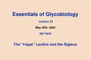
I-Cell Disease“Inclusion Cell Disease” Fibroblasts
Fibroblasts from a human genetic disease with prominent "inclusion bodies" in cells ("I-cell disease") lack not one, but almost all lysosomal enzymes. In I-cells, all the enzymes are actually being made, but are almost completely secreted into the medium. Hickman and Neufeld: I-cells could take up the "high-uptake" enzymes from normal cells, but the enzymes secreted by I-cells not taken up by other cells. I-Cell Disease
Hypothesis: I-cell disease resulted from a failure to add a “common recognition marker” present on all lysosomal enzymes I-Cell Disease
Complex -type glycans Endo-beta-N-acetyl- glucosaminidase H (Endo-H) Hybrid-type glycans Major Steps in the Biosynthesis of N-glycanson Glycoproteins(including Lysosomal Enzymes) High mannose-type glycans
Uptake of "high-uptake" lysosomal enzymes specifically blocked by mannose 6-phosphate (M6P) and its stereoisomer fructose-1-phosphate. Millimolar concentrations required, but similar concentrations of other sugars and sugar phosphates had no comparable effect. Since Man residues occur on high mannose-type N-glycans, it was predicted that these might be phosphorylated specifically on lysosomal enzymes. Structural Nature of the “High-Uptake” Marker
Confirmed by alkaline phosphatase treatment, which abolished "high-uptake" activity, and by tunicamycin treatment, which blocked N-glycosylation, and caused secretion of lysosomal enzymes from cells. M6P directly shown to be present in "high-uptake" forms of lysosomal enzymes and on EndoH-sensitive N-glycans from these enzymes Structural Nature of the “High-Uptake” Marker
Endo-H sensitive N-glycans of Lysosomal Enzymes Contain “Blocked” Phosphate residues X-P- Man-(N-glycan)-Lysosomal enzyme Endo H X-P- Man-(N-glycan) Mild Acid X + P- Man-(N-glycan) Alkaline Phosphatase P + Man-(N-glycan) X = GlcNAc!
Enzymatic Steps in the Biosynthesis of the High-uptake Marker Uridine-P-32P-a[6-3H]GlcNAc + Mana1-(N-glycan)-Lysosomal enzyme “Phosphotransferase” Uridine-P + [6-3H]GlcNAca1-32P-6-Mana1-(N-glycan)- Lysosomal enzyme “Uncovering Enzyme” [6-3H]GlcNAc + 32P-6-Mana1-(N-glycan)- Lysosomal enzyme Phosphatase 32P + Mana1-(N-glycan)- Lysosomal enzyme
MANNOSE-6-P-GlcNAc MANNOSE-6-P MANNOSE-6-P RECEPTOR (S) GlcNAc-PHOSPHO- TRANSFERASE PHOSPHODIESTER GLYCOSIDASE DEFECT IN I-CELL DISEASE MANNOSE MANNOSE-6-P LOW pH ACID PHOSPHATASE LYSOSOME ENDOSOMAL COMPARTMENT Mannose 6-phosphate pathway for trafficking of lysosomal enzymes GOLGI APPARATUS ENDOPLASMIC RETICULUM MANNOSE N-LINKED SUGAR CHAIN LYSOSOMAL ENZYME
Pseudo-Hurler Polydystrophy (Mucolipidosis III)A Milder version of I-cell Disease
Nature of the Defect in a Variant form of Mucolipidosis III Failure to Recognize Lysosomal Enzymes as Special Substrates?
Not explained by similarities in primary polypeptide sequences Denatured lysosomal enzymes loose specialized acceptor activity Features of secondary or tertiary structure crucial Sequence "swapping" between cathepsin D (M6P+) and pepsinogen (M6P-) scattered basic residues critical (particularly lysines) two regions of the cathepsin D amino lobe are involved these cooperate with a recognition element in the carboxyl lobe Structural Basis for Recognition of Lysosomal Enzymes by GlcNAc Phosphotransferase
Studies with other enzymes confirm general model: scattered basic residues + adjacent surface loops How does catalytic reach of GlcNAc-phosphotransferase extend to widely spaced N-glycans on a lysosomal hydrolase target? Different N-glycans on the same enzyme get different degrees of phosphorylation, based on how far away they are from the recognition patch(es) Structural Basis for Recognition of Lysosomal Enzymes by GlcNAc Phosphotransferase
Catalyzes initial step in synthesis of the mannose 6-phosphate determinant Partially purified by chromatography and used to generate a panel of murine monoclonal antibodies Monoclonal antibody coupled to a solid support and used to immunopurify the enzyme ~480,000-fold to apparent homogeneity Purification of UDP-N-acetylglucosamine:lysosomal-enzyme N-acetylglucosamine-1-phosphotransferase (GlcNAc-phosphotransferase) 1996
Subunit structure: 540,000-Da complex of disulfide-linked homodimers of 166,000- and 51,000-Da subunits and two identical, noncovalently associated 56,000-Da subunits Properties essentially same as those originally described for impure enzyme Human cDNA and genomic clones reported in 2000 Purification of UDP-N-acetylglucosamine:lysosomal-enzyme N-acetylglucosamine-1-phosphotransferase (GlcNAc-phosphotransferase) 1996
Catalyzes second step in the synthesis of mannose 6-phosphate determinant of lysosomal enzymes Partially purified preparation used to generate a panel of murine monoclonal antibodies. Monoclonal antibody coupled to a solid support and used to immunopurify the bovine liver enzyme ~670,000-fold in two steps to apparent homogeneity N-Acetylglucosamine-1-phosphodiester alpha-N-Acetylglucosaminidase(“Uncovering Enzyme” or Phosphodiester alpha-GlcNAcase)
Purified enzyme has similar properties to original one Subunit structure: complex of 4 identical subunits arranged as two disulfide-linked homodimers - a type I membrane-spanning glycoprotein with amino terminus in lumen of Golgi apparatus Human cDNA and mouse genomic DNA clones isolated in 1999 N-Acetylglucosamine-1-phosphodiester alpha-N-Acetylglucosaminidase(“Uncovering Enzyme” or Phosphodiester alpha-GlcNAcase)
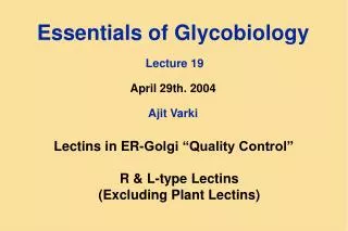
Cell/Tissue with Binding Sites for M6P ligands Non-ionic Detergent Extract Apply to Affinity Column Under physiological conditions M6P M6P Wash well With buffer SDS-PAGE Isolation of the Mannose-6-Phosphate Receptors(M6PRs, P-type lectins) Ca++/Mg++ Reapply To column Reproduce Elution specificity Dialyze 1mM Glc6P & discard Elute With? 1mM Man6P
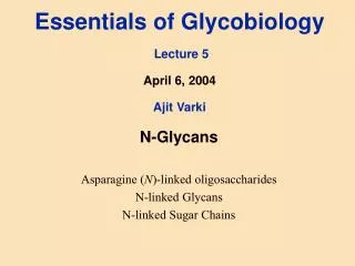
Monomer 1 Monomer 2 Ribbon diagram of the CD-MPR (Roberts et al., Cell 93:639-648, 1998) Man-6-P Man-6-P
NO 1 = Golgi Mannosidase I Complex and hybrid-type glycans 2 = GlcNAc Phosphotransferase 3 = GlcNAc Transferase I 4 = Phosphodiester glycosidase * * 5 = Galactosyltransferase -P- -P * * +/- 6 = Sialyltransferase(s) 4 E 1 * -P- * 5,6 * + -P * 4 D 1,2,3 1 * * P- -P -P- -P- * * -P- * +++ 4 2 2 A C B BINDING TO MPRs Biosynthesis of Phosphorylated N-glycans NO NO +/-
Targeted disruption of CD-MPR gene: normal or only slightly elevated levels of lysosomal enzymes, otherwise normal phenotype. However: thymocytes from homozygous CD-MPR null mice or primary cultures of fibroblasts show increase in lysosomal enzyme secretion Other glycan-specific endocytotic receptors (mannose-specific receptor of macrophages or asiaoglycoprotein receptor of hepatocytes) provide in vivo compensation? Genetic Defects in the Mannose 6-Phosphate receptors
Mouse CI-MPR is part of the naturally ocurring Tme locus, a maternally imprinted region of chromosome 17 (i.e. expressed only from the maternal chromosome). Mice that inherit a deleted Tme locus from their mother die at day 15 of gestation. Lethality due to lack of CI-MPR - proven by induced disruption of gene. Maternal inheritance of null allele generally lethal by birth and mutants about 30% larger in size. Size phenotype probably caused by excess IGF-II, because introduction of an IGF-II null allele rescued the mutant mice. Mutant mice also have organ and skeletal abnormalities Genetic Defects in the Mannose 6-Phosphate receptors
Fibroblasts prepared from embryos that lack one or both receptors. Fibroblasts lacking only one receptor showed a partial impairment in enzyme sorting. Fibroblasts lacking both receptors show massive missorting of multiple lysosomal enzymes and accumulated undigested material in their endocytic compartments. Thus, both receptors are required for efficient intracellular targeting of enzymes. Genetic Defects in the Mannose 6-Phosphate receptors
Comparison of phosphorylated proteins secreted by different cell types indicates that the two MPRs interact preferentially with different subgroups of hydrolases. Confirmed by in vitro studies using different enzymes and cell types Heterogeneity of phosphomannosyl recognition marker within a single enzyme and amongst different enzymes explains evolution of two MPRs with complementary binding properties. Together with factors such as the number, compartmental localization, properties and availability of receptors, the endosomal pH, and concentration of divalent cations, there is much flexibility in this trafficking mechanism Genetic Defects in the Mannose 6-Phosphate receptors
Lysosomal enzymes successfully targetted in Saccharomyces, Trypanosoma and Dictyostelium, without any identifiable MPRs. D.discoideum produces a methyl-phosphomannose sequence on some lysosomal enzymes that can be recognized by the mammalian CI-MPR (not the CD-MPR ). There is also a GlcNAc phosphotransferase that recognizes lpha1-2 linked Man residues, but it is not specific for lysosomal enzymes. Acanthamoeba produces a phosphotransferase that does show specific recognition of mammalian lysosomal enzymes. Evolutionary origins of the MPR system
Although some of these organisms show evidence for an “uncovering” enzyme, no definable MPR has been found. A CI-MPR receptor was recently identified in a mollusc. Evolutionary divergence point at which complete MPR system emerged has yet to be definitively identified. Evolutionary origins of the MPR system
In I-cell disease, some cells and tissues (e.g. liver, B-lymphoblast lines and circulating granulocytes) have essentially normal levels of lysosomal enzymes. Two soluble lysosomal enzymes, acid phosphatase and ß-glucocerebrosidase are not at all affected in their distribution even in I-cell disease fibroblasts. Acid phosphatase begins life as a membrane-bound protein, and once in the lysosome, it is proteolytically cleaved to generate the mature soluble form Alternate Pathways for Trafficking of Lysosomal Enzymes
Glucocerebrosidase is soluble, but membrane associated, does not show phosphorylation, and is targetted to lysosomes independent of this pathway. Is the M6P pathway for trafficking of lysosomal enzymes a specialized form of targetting, superimposed upon some other basic mechanisms that remain undefined? Note: Integral membrane proteins of lysosome such the lysosomal membrane proteins also do not require the phosphomannosyl recognition pathway for trafficking to lysosomes. They utilize motifs in their cytosolic tails similar to those of the MPRs Alternate Pathways for Trafficking of Lysosomal Enzymes
N.Dahms Man-6-P-Containing Proteins Man-6-P Ligand Consequence of Binding to MPR INTRACELLULAR targeted to lysosomes, lysosome biogenesis internalized cytokine degraded in lysosomes internalized growth inhibition proteolytic activation growth inhibition proteolytic activation cardiac angiotensin I internalized induction of apoptosis internalized enhanced T cell migration/activation induction of angiogenesis/endothelial cell migration internalized enhancement of viral entry internalized enhancement of viral entry Lysosomal enzymes Leukemia inhibitory factor CREG TGF- precursor Renin precursor Granzyme B CD26 Proliferin Herpes simplex virus gD Varicella-zoster virus gI EXTRACELLULAR
internalized mitogen degraded in lysosomes internalized degraded in lysosomes modulate uPAR’s interaction with integrins and vitronectin proteolytic activation generation of plasmin mediates growth inhibitory effects of retinoic acid Insulin-like growth factor II (IGF-II) Urokinase-type plasminogen activator receptor (uPAR) Plasminogen Retinoic acid Non-Man-6-P-Containing Ligands of CI-MPR Ligand Consequence of Binding to CI-MPR EXTRACELLULAR N.Dahms
Domains 1-15 vs Domains 1-15 Domains 1-15 vs CD-MPR 16-38% identity 14-28% identity 43aa fibronectin type II-like insert N-glycosylation site Palmitoylation M6P M6P P P 300kDa CI-MPR Two Mannose 6-Phosphate Receptors Plasminogen 1 1 uPAR 1 2 2 2 M6P 3 3 3 M6P----X----M6P 4 4 4 5 5 5 6 6 6 7 7 7 8 8 8 M6P 9 9 9 M6P----X----M6P 10 10 10 11 11 11 IGF-II 12 12 12 13 13 13 14 14 14 15 15 15 Retinoic acid? Cytosol P P P 46kDa CD-MPR P P P cation-dependent cation-independent (IGF-II Receptor) N.Dahms
N.Dahms TGF-β active CI-MPR = Tumor suppressor 1) Decreases serum levels of mitogen IGF-II 2) Activates growth inhibitors TGF-, CREG IGF-II IGF-II Multifunctional 300kDa CI-MPR plasminogen plasmin Cell motility uPA Growth inhibitory effects CI-MPR Vitronectin M6P Integrins uPAR retinoic acid inactive TGF-β Plasma membrane Cytosol Growth inhibitory effects Signaling Cascades Degradation IGF-II = Insulin-Like Growth Factor II uPAR = Urokinase-Type Plasminogen Activator Receptor