Download
1 / 69
740 likes | 1.26k Views
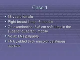
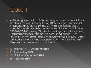
Case 1. •25 y.o. WF college student referred for anemia with low iron. MCV 72, Retic 0.5% 6 mo. ago 5 K times dropped to 21 min. • Fatigued (working too hard at school) • No menstrual period in 1 year, not pregnant •Under treatment for anorexia

E N D
Case 1 •25 y.o. WF college student referred for anemia with low iron. MCV 72, Retic 0.5% 6 mo. ago 5 K times dropped to 21 min. • Fatigued (working too hard at school) • No menstrual period in 1 year, not pregnant •Under treatment for anorexia •Pregnant ~ 3 years ago, no iron replacement
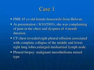
Iron Deficiency Hypochromia Pencil Cells
Who Loses How Much Iron Daily? • Adult Male, Menopausal female 0.5-1 mg • Menstruating female 1-2 mg • Pregnant female 1.5-3 mg • Children 1 mg • Female 12-15 yr 1.6-2.6 mg Compare to blood loss 250 mg/500 ml
Case 2 A 42 y.o. man presents with back pain (2 weeks), Fever to 102. Leg weakness •Fell prior to coming in, down for 4 hours in the bathroom and couldn’t get up. •Weight = 420 pounds. •Urine red. •Hgb is 9.2, MCV is 74. •Iron/TIBC is 15/200 (Sat ~8%) •Blood Culture + for Staph species
Does This Body Have Iron?Look in the Bone Marrow (Normal) Macrophage has lots of iron. Won’t share with RBC. Selfish Macrophage? Anemia of Chronic Disease (~1/3 are microcytic)
Iron Stores Too Much None
MCV for Iron LackJGIM 1992;7:145-153 MCV Likelihood Ratio >90 0.29 85-90 0.76 80-85 0.91 75-80 1.0 70-75 3.3 <70 12.47
Transferrin Saturation ACD vs Iron Deficiency Transferrin Saturation LR >50% 0.15 30-50% 0.43 20-30% 0.52 10-20% 0.81 5-10% 2.54 <5% 10.46
Serum Ferritin (ACD vs Fe-Lack) Ferritin Level LR >100 0.12 45-100 0.54 35-45 1.83 25-35 2.54 15-25 8.83 <15 51.85
30 100 12
Serum Transferrin ReceptorIncreased in iron lack False Positives: Hemolysis, Megaloblastic Anemia
Tests for Iron Deficiency 100 50 0 MCV Sat’n Ferritin True Positive ROC Curve Guyatt 0 50 100 False Positive
Tests for Iron Deficiency 100 50 0 TfR/Ferritin TfR Ferritin True Positive ROC Curve Punnonen 0 50 100 False Positive
Diagnosis of Iron Lack Clinical Suspicion Ferritin <12 - iron deficiency >100 probably not In Between Gold Standard
Therapeutic Trial Oral Iron Max Retics at 10-14 days Hemoglobin rise 2 gm in 1 month OR 0.25 gm/d after peak retic
Case 3 •64 y.o. WM is followed for chronic lymphocytic leukemia. •Has noted increasing fatigue. He has been chronically anemic at ~Hgb. 10.8. (with all normal indices, including MCV of 95 at last visit) •Anemia increased - Hgb 8.2, microcytic & hypochromic. Retic count 1%. •Fe/TIBC = 15/380 (saturation = 4%), Ferritin 8
How Far Should We Go ToInvestigate Blood Loss Source GOLD STANDARD : Upper and Lower Endoscopy in men and post-menopausal women WHY? 3/4 of patients will have a lesion.
Evaluation of GI Tract About 2/3 of IDA patients are symptomatic Among asymptomatic, 1/2 will have lesion (44% guaiac negative) Wilcox et al., Am J Med 1997, 103:405
Endoscopic Evaluation of IDA Hardwick et al. Br J Surg 1997; 84:1725 89 patients with IDA UGI cause 25 LGI cause 24 Both UGI and LGI 26
What Did They Have? Wilcox et al., Am J Med 1997, 103:405 Among 52 asymptomatic patients with IDA: Colon cancer 11 (21%) Vascular ectasia 9 (17%) Gastric carcinoma 1 Colonic Ulcer 1 Colonic Polyp 1
Bad Things Found Hardwick et al. Br J Surg 1997; 84:1725 UGI Carcinomas 9 UGI Lymphomas 2 LGI Carcinomas 17 Crohn’s 3 Celiac 2 Ulcerative Colitis 5
Some Special Points 1. Malignancies are not always guaiac + (3 of 9 negative in Kepczyk study) 2. Bleeding on NSAID does not preclude need for endoscopy 3. Small bowel biopsy will identify some celiac disease (if not your routine, consider if patient refractory to oral iron)
Iron Lack vs. ACD • Ferritin is 1st test for Iron Lack vs. ACD • Serum Transferrin Receptor or Therapy Trial is next • W/U of iron lack includes Upper and Lower Endoscopy. • Small Bowel Bx and/or anti-endomysial, anti-gliadin • Endoscope premenopausal women: if guaiac +, severe anemia, GI symptoms, non-correction with iron
Case 4 A 73 y.o. WM of Swedish heritage presents with fatigue, depression and anemia(Hb 7.0) •Bili 2.4, LDH 1500 •He has poor appetite, weight loss of 15 # •W/U so far has included negative stool quaiacs, GI series, upper and lower endoscopies, liver scan. •Physical exam: Flattened affect, pale lemon yellow skin, subtle scleral icterus •Neurological: absent vibration and position sense (feet) •MCV 125, Retic 1.8%
Marrow: Nuclei are losing the race to maturity
What is Megaloblastosis? • Big cells (macrocytosis) • Impaired nuclear maturation. PRODUCTION PROBLEM Low Retic Count
Clues to Megaloblastosis Size: MCV > 120 usually megloblastic Macro-ovalocytes Hypersegmentation
Macrocytosis Plasma Lipids Lipid-loaded Reticulocytosis Liver Disease Hypothyroidism
Diagnosis of B12 Deficiency Serum B12: >300 – unlikely (1-5%) <200 – likely (95+% specificity) 200-300 – possible (check Methyl Malonic Acid - MMA)
Pernicious Anemia Autoimmune: Antibodies to parietal cells & IF. Deficient IF leads to low B12 More common in elderly, northern European
Problems Associated with PA Neurological Problems: Posterior Columns Lose Position and Vibratory Depression, lability Megaloblastic Madness Other autoimmune problems: thyroid, adrenal, vitiligo (anti-melanocyte)
Ways to Get B12 DeficiencyOther Than PA Walleye Souchi: Diphyllobothrium latum (fish tapeworm) is indigenous in Great Lakes region. Competes for B12. Inflammatory Bowel Disease (ileum) Gastric resection Blind Loop: Bacterial overgrowth
Folate Deficiency • Dietary cause more likely than with B12 due to lesser stores. • More typically deficient in diet of alcoholics • Malabsorbed in sprue, Crohn’s • Excessive use in hemolytic anemia, exfoliative dermatitis, pregnancy, prematurity.
Folate and Alcohol • Serum folate drops abruptly in four days with alcohol abuse (decrease absorption and enterohepatic circulation) • With dietary deficiency, 4-5 months to develop folate-lack megaloblastosis. • If add alcohol abuse to poor diet, takes 5 to 10 weeks. • Case finding with large MCV: ~10-20% will be alcoholic
Lab Tests for Folate Lack • Serum Folate can increase to normal with a single hospital meal, BUT • If <2 is fairly diagnostic • If >4, fairly reliable to rule out.
If Serum Folate 2 to 4 Check RBC Folate Is not rapidly altered with diet. Reflects stores. Homocysteine will also be high (in both folate and B12 deficiency)
If megaloblastic, but folate and B12 are in “iffy” range (If folate >4 and B12 >300, no more testing) Can check both homocysteine and MMA: B12 deficiency Folate MMA 98% 12% homocysteine 96% 91% Savage et al., Am J Med 1994; 96:239
Case 5 32 y.o female with H/O Stage III A Hodgkin’s 8 years ago presents with fever to 104, shaking chills, dark urine. Very ill with BP 70/20. On no meds. Hemoglobin 8, Reticulocytes 7%. WBC 13,000 with shift to left. Platelets 20,000. Urinalysis + for large blood by dipstick, no red cells on microscopic. Blood culture + for Streptococcus pneumonia.
How Do We Decide There is Hemolysis? • Measure of turnover? • Damage to red cells? • Intra vs Extravascular
How Many Retics is Too Many? With anemia, need 4-5% to say is destructive Mean in autoimmune hemolysis ~9% Absolute retics should be high (RBC number X % retics) Normal = 5 million X 1-2% =50 to 100 thousand
Increased Retics Can Mean • Hemolysis • Blood Loss • Replenishment of “building blocks” like iron, folate, etc.
Does Hemolysis Always Cause Reticulocytosis? Not in the following: • Chronic hemolysis and ran out of folate • Chronic hemolysis and developed a viral infection which shut down marrow production (Owren’s Crisis) • Coombs positive hemolysis with antibodies also attacking marrow precursors (10-20%)
Intravascular • Only THREE tests which are specific: • Plasma Hemoglobin • Urine Hemoglobin • Urine Hemosiderin
Case 6 48 yo F presents with sun sensitivity (face) Fatigue, intermittent joint pains Hemoglobin 8. MCV 105. Retic 15%. WBC 2500, platelets 105,000.
Direct Antiglobulin Test – Coombs Polyvalent Ab Against Ig, C’ RBC from patient, coated with Ig or C’ Agglutination - + Test Elute – IgG or C’