Download
1 / 198
1.99k likes | 2.24k Views
Unit 2—Principles of Support & Movement. Dr. Achilly. Part 1: Bone tissue. “Concepts” chapter 8. Functions . Bone tissue serves many functions: Support—framework for body & attachment point for muscles. Protection—for soft internal organs

E N D
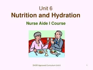
Unit 2—Principles of Support & Movement Dr. Achilly
Part 1: Bone tissue “Concepts” chapter 8
Functions • Bone tissue serves many functions: • Support—framework for body & attachment point for muscles. • Protection—for soft internal organs • Assists movement—muscles need to be attached to stable structures in order to produce movement.
Functions • Mineral reserve—stores calcium & phosphorus to help maintain homeostasis in their blood levels. • Blood cell production—some bones have red marrow which produces red & white blood cells & platelets. • Triglyceride storage—yellow bone marrow stores these for energy reserve.
Structure • Bone is made up of several tissues: • Osseous • Cartilage (fibrous connective tissue) • Connective (for binding and support) • Epithelium (covering) • Adipose (fat) • Nervous • Each bone is considered an organ & each is continually remodeling.
Macroscopic Structure • In a typical long bone (femur, humerus) you’ll find: • Diaphysis—shaft or long main part of bone. • Epiphyses(pl)—distal and proximal ends • Metaphyses(pl)—regions where diaphysis joins each epiphysis (sing). When still growing this region contains an epiphyseal plate —an area of cartilage that allows for elongation. This plate is replaced by an epiphyseal line when the bone stops growing.
Macroscopic Structure • Articular cartilage—thin layer that covers the epiphysis when the bone articulates (forms a joint) with another. This hyaline cartilage is slippery and absorbs shock. • Periosteum—sheath that surrounds the bone. These cells are bone-forming & add to thickness of bone. They also protect, nourish, repair & serve as attachment for tendons & ligaments. Perforating Sharpey’s fibers act like thumbtacks to hold the periosteum to the bone.
Macroscopic Structure • Medullary cavity—aka marrow. Space within the diaphysis. • Endosteum—thin membrane that lines the medullary cavity.
Microscopic Structure • Not solid. Bone calcifies when crystals of hydroxyapatite (calcium, magnesium, potassium…) form around a framework of collagen. • Bone is hard & flexible. • Composed of 4 types of cells: • Osteogenic— divide & develop into osteoblasts. • Osteoblasts—bone building. Secrete extracellular matrix & initiate calcification. Become osteocytes. • Osteocytes—mature bone cells.
Microscopic Structure • Osteoclasts—concentrated in endosteum. Face bone surface. Release enzymes that break down bone matrix. Called resorption.
Microscopic Structure • Bone has spaces that act as storage areas, canals for blood/nerve supply. • Classified based on size/amount of spaces.
Microscopic Structure • Compact bone • Few spaces, very strong. Found beneath periosteum and makes up most of the diaphysis. • Blood/lymphatic vessels & nerves penetrate bone thru perforating canals (Volkmann’s). Connect to central canals called haversian canals. • Orientation of these structural units (osteons) can change with age, stress, injury.
Microscopic Structure • Spongy bone • No regular osteons. Irregular lattice of trabeculae instead. • Lighter • Makes up more areas of flat bones and the epiphyses of long bones. • Orient along lines of stress. Helps bones transfer stress w/o breaking. • Red marrow located here.
Microscopic structure • Bones are well supplied with arteries, veins & nerves.
Bone formation • Intramembranous ossification • Flat bones in embryo form this way. • Osteogenic cells cluster where bone will form. Extracellular matrix is secreted to begin calcification. • Trabeculae and periosteum develop.
Bone formation • Endochondral ossification • Bone replaces a cartilage “scaffold.” • Seen in long bones. • Starts as primary ossification center develops in diaphysis. Ossification spreads inward. Medulla (marrow) eventually forms. • Secondary ossification center develops at birth in the epiphyses. Ossification spreads outward.
Bone growth • Increase length • Diaphysis lengthens b/c of activity at epiphyseal plate. New chondrocytes (cartilage forming cells) form on epiphyseal side while old chondrocytes on diaphyseal side are replaced by bone. • Damage to epiphyseal plate can cause bone to not reach its normal length.
Dark line indicates epiphyseal plate Salter-Harris IV Fracture . White arrow points to metaphyseal fracture and yellow arrow to a fracture of the distal tibial epiphysis in this Salter-Harris IV fracture of the ankle.
Bone growth • Increase in thickness • Bone grows outward from periosteum. Cells here become osteoblasts and build new osteons. • Bones are always being remodeled. Bones subjected to heavy loads will grow thicker. • Remodeling is dependent upon intake of nutrients and presence of hormones. • Minerals: calcium, phosphorus, iron, manganese, fluoride, magnesium • Vitamins: C, K, B12, A • Hormones: growth factors, androgens
Bone and homeostasis • Bone is a major calcium reservoir.
Cartilage • In order to bring 2 bones together in a joint, cartilage is needed. • Made of collagen fibers for strength & chondroitin sulfate for resilience. • Three types: • Hyaline—found at ends of bones, most abundant, but weakest. Reduces friction and absorbs shock. • Fibrocartilage—found in intervertebral discs, menisci. Strongest. Reduces friction. • Elastic—Found in larynx, ear. Gives support and maintains shape.
Part 2: Axial skeleton “Concepts” chapter 9
Divisions of the skeletal system • Axial • Bones that lie on the longitudinal axis of the body. • Skull • Hyoid • Auditory ossicles • Vertebral column • Thorax (sternum & ribs)
Divisions of the skeletal system • Appendicular • Pectoral (shoulder) girdle = clavicle & scapula • Upper limb = humerus, ulna, radius, carpals, metacarpals, phalanges • Pelvic girdle = coxal/innominate bones • Lower limb = femur, patella, fibula, tibia, tarsals, metatarsals, phalanges
Bone types • Long • Greater length than width (femur) • Short • Nearly equal in length and width (carpals) • Flat • Flat & thin (cranial, sternum) • Irregular • Complex shapes (vertebrae, calcaneus) • Sesamoid bones • Shaped like sesame seeds (patella)
Surface markings • In general there are 2 major surface markings on bones of the body: • Depressions (aka foramen, fossa, sulcus)—allow nerves, vessels or tendons to pass thru bones. • Processes (aka condyle, facet, crest, trochanter, tuberosity)—help to form joints or points for ligament attachment.
Skull • Made of 22 bones grouped into two categories: • Cranial • Facial
Skull • Cranial bones (8) encase brain • Frontal bone • 2 parietal bones • 2 temporal bones • Occipital bone • Sphenoid bone • Ethmoid bone
Skull • Facial bones • 2 nasal bones • 2 maxillae • 2 zygomatic bones • Mandible • 2 lacrimal bones • 2 palatine bones • 2 inferior nasal conchae • vomer
Skull practical practice • Nice link: • http://www.gwc.maricopa.edu/class/bio201/skull/antskul.htm
Skull • Bones of the skull continue to grow until about age 14. • There is space between the sutures to allow for growth. • Babies have fontanels (soft spots) where ossification of the bone has not occurred.
Hyoid bone • Unique because it doesn’t articulate with any other bone. • Suspended from styloid processes of temporal bones by ligament & muscles. • Helps support & anchor tongue & its muscles.
Vertebral column • a.k.a. spine • Consists of bone, connective tissue • Surrounds & protects the spinal cord. • Adult spine consists of 26 vertebrae • 7 cervical • 12 thoracic • 5 lumbar • 1 sacrum • 1 coccyx
Vertebral column • Four normal curves in adult spine: • Cervical lordosis • Thoracic kyphosis • Lumbar lordosis • Sacral kyphosis
Vertebral column • Between each vertebrae is an intervertebral disc. • Outer fibrocartilage ring • Inner spongy, soft tissue • Help to form strong joints & absorb shock.
Vertebral column • Vertebrae will look different depending upon the area of the spine. • They all have 3 common parts: • Body • Vertebral arch • Processes
Vertebral column • The first 2 cervical vertebrae are unique. • Atlas (C1) has no body, articulates with cranium, has large transverse processes(TP’s). • Axis (C2) has no body, but has peg-like structure called the dens. • Allows for rotation & nodding motion of cranium.