Download
1 / 28
280 likes | 638 Views
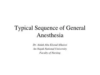
Typical Sequence of General Anesthesia. Dr. Aidah Abu Elsoud Alkaissi An-Najah National University Faculty of Nursing. Size PLAIN. Size CUFFED. 2.5 mm. 4.5 mm. 3.0 mm. 5.0 mm. 3.5 mm. 5.5 mm. 4.0 mm. 6.0 mm. 4.5 mm. 6.5 mm. 7.0 mm. 7.5 mm. 8.0 mm. 8.5 mm. 9.0 mm.

E N D
Typical Sequence of General Anesthesia Dr. Aidah Abu Elsoud Alkaissi An-Najah National University Faculty of Nursing
Size PLAIN Size CUFFED 2.5 mm 4.5 mm 3.0 mm 5.0 mm 3.5 mm 5.5 mm 4.0 mm 6.0 mm 4.5 mm 6.5 mm 7.0 mm 7.5 mm 8.0 mm 8.5 mm 9.0 mm Size of the tubesplain and cuffed
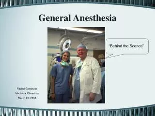
Typical Sequence of General Anesthesia • Pat is identified • Checked for consent • Result test & Dx studies • I.v infusion startedpreop. Or on arrival • All drugs are given i.v. • Intraoperative monitoring
Typical Sequence of General Anesthesia • At induction- preoxygination with 100% O2, 3-5 minutes through a mask- permits washout of most of the gaseous nitrogen from the body and provides a large reserve of oxygen in the lungs • A test dose of the induction agent(50 mg thiopental) is given to check for exagerated response • If succinylcholine will be used for intubation then a small pretreatment dose of a nondepolarizing muscle relaxant (0.5 mg of pancuronium) is given • When given 5 min before succinylcholine, this small dose is sufficient to block most of the muscle fasciculations seen with succinylcholine and decrease post.op myalgias
Typical Sequence of General Anesthesia • To induce anesthesia, and move pat rapidly to stage 3, a short acting barbiturate as thiopental (2-6 mg/kg) is given • When pat becomes apneic and the eyelid reflex is gone, the airway is checked for patency by ventilating the pat with a mask • Depending on several Factors such as an adequate airway and the type and duration of surgery, oxygen and anesthetic gases may be delivered to a spontaneously breathing pat via a mask that is held in place with a head strap • Positioning of the head and oral or nasal airway used to maintain a patent airway • If mask anesthesia is not suitable, then endotracheal tube may used to facilitate ventilation or to protect the airway from aspiration
Typical Sequence of General Anesthesia • An intubating dose of a muscle relaxant such as succinylcholine (1-1.5 mg/kg) is given • Muscle relaxant causes temporary paralysis of the jaw and diaphragmatic muscle as well as other skeletal muscles • To facilitate intubation, a laryngoscope held in the left hand is inserted into the right side of the mouth and then moved to the midline ”sweeping” (taking in or moving over) tounge to the left • The endotracheal tube is introduced on the right side of the mouth and gently inserted into the trachea so that the cuff is a cm below the vocal cords • The cuff is inflated just enough to occlude any air passage with the peak pressures used for ventilation
Typical Sequence of General Anesthesia • Correct location of the endotracheal tube is verified by lisening for bilaterally equal breath sounds with a stethoscope, • by absence of sounds over the stomach and by an appropriate level and waveform of ETCO2 • By symmetrical movement of the thorax with positive pressure ventilation. look at the chest for expansion with each breath • By condensation of moisture from expired air in the endotracheal tube and breathing circuit • ausculatate the chest for breath sounds • An appropriate level and waveform of ETCO. check the capnoraphic tracing on the monitor to ensure end tidal CO2
Typical Sequence of General Anesthesia • The vocal cords are the narrowest portion of n adult traches, the smallest portion of a child´s airway is below the vacal cord • Thereore uncuffed endotracheal tubes are used for children because the internal diameter of cuffed tubes in these small sizes would have too much resistance to ventilation and could easily become obstructed • After the paralysis from the succinylchiline has worn off, the pat may be allowed to breath spontaneously with intermittent assistance, or additional muscle relaxant may be given and the ventilation controlled either manually or mechanically
Typical Sequence of General Anesthesia • If the procedure is an emergency or the pat is at risk for aspiration (for example, in cases of intestinal obstruction, full stomach, hiatal hernia) • The anesthesiologist may undertake rapid sequence induction • Standing on the patient´s right side • The perioperative nurse must be ready to assisst as necessary by applying downward pressures on the cricoid cartilage with the thumb and index finger of the right hand while the left hand is plaved under the patient´s neck for stability • The cricoid cartilage is the only complete ring in the trachea, and downward pressure occludes the esophagus which lies immediately posterior
Typical Sequence of General Anesthesia • If, after assessment of the patient´s airway, the anesthesiologist feels rapid intubation of trachea may be difficult, an awake intubation, using a fiber optic bronchoscope or other techniques, may be chosen • Maintenance of anesthesia can be accomplished with either intravenous or inhalational anesthetic techniques or a combination of both with or without additional muscle relaxant • The anesthesiologist suctions the oropharynx to decrease the risk of aspiration or laryngospasm following extuv´bation, reverse any residual paralysis from the nondepolarizing muscle relaxant with an anticholinesterase such as neostigmine and an anticholinergic such as atropine and allows wash out of nitrous oxide and volatile anesthetic agents by giving 100% oxygen for several minutes before extubation • The pat is then transported to the PACU to a waken from the anesthetic experience
Typical Sequence of General Anesthesia • Untoward events that can occur with general anesthesia include:hypoxia, respiratory, cardiovascular,renal dysfunction • Hypotension • Hypertension • Imbalance of fluids or electrolytes • Residual muscle paralysis • Neurologic problems • Malignant hyperthermia
Prior to intubation, • always check equipment and make sure everything you might need is not only within your reach, but also properly working. • If in the operating room, a complete check of the anesthesia equipment at the start of each day as well as a modified check before each new case is imperative. • If in the emergency room or the hospital wards, make sure you know where all of your equipment is and, also, that you have the necessary resources to support the patient once intubated.
Prior to positioning the patient: • Make sure that your laryngoscope is locked into position and that the incandescent light on the blade tip functions. • Also make sure that you have several alternate blades available in case the one you have chosen does not allow for visualization of the cords. • Examine the endotracheal tube. Make sure that the cuff inflates by using a 10-mL syringe to inflate the cuff and then detach the syringe to ensure that the cuff pressure is maintained. Be sure to deflate deflate the cuff after testing it. • Attach the connector to the proximal end of the tube. Push it in as far as possible to lessen the likelihood of disconnection. • If you are going to use a stylet, it should be inserted into the ET tube and bent to resemble a hockey stic • using a stylet, one should be within easy access in case the intubation proves to be more difficult than anticipated. • Ensure a functioning suction unit to clear the airway in case of unexpected blood, emesis or secretions. • Ensure that you have tape within your reach to secure the tube once it is in place
positioning • Proper patient positioning can be the difference between a successful and failed intubation. • The patient’s head should be level with the physician’s xiphoid process. • To achieve the sniff position (which allows for optimal visualization of the glottic opening), elevate the patient’s head and extend the atlanto-occipital joint. This can be achieved by sliding your free hand (right hand if you are right handed, left hand if you are left handed) beneath the patient’s head and gently lifting it up and towards you. Or, you can gently position the chin up and mouth open before attemting laryngoscopy. • The "scissor technique" can also be used to further open the patients mouth. Cross your right forefinger and thumb and insert into the right side of the patient's mouth. Apply pressure to the upper teeth with your forefinger and the lower teeth with your thumb to open the mouth. Be sure to position your hands so as NOT to obstruct your view.
OTHER WAYS TO INTUBATE • Nasotracheal Intubation: Nasal intubation is similar to oral intubation except that the ETT is advanced through the nose into the oropharynx before laryngoscopy. • If the patient is awake, local anesthetic drops and nerve blocks can be used. • A lubricated ETT is introduced along the floor of the nose, below the inferior nasal turbinate, perpendicular to the face. • Often, a nasopharyngeal airway can be used. The tube is advanced until it can be visualized in the oropharynx. Via laryngoscopy, the tube is then advanced in between the abducted vocal cords. • Nasal instrumentation (with ETTs, NPOs, or nasal catheters) is contraindicated in all patients with severe midfacial trauma.
Flexible Fiberoptic Bronchoscopy: • Laryngoscopy may be contraindicated in a patient who requires intubation and mechanical ventilation. This is often the case in trauma patients who may have an unstable cervical spine or in patients with poor range of motion of the temporo-mandibular joint. In such patients, flexible fiberoptic bronchoscopy allows for indirect visualization of the larynx. The endoscope is introduced through the mouth or nose. Once anatomic structures are recognized, and the larynx or trachea are entered under direct visualization
COMPLICATIONS OF INTUBATION • Complications of laryngoscopy and intubation are most frequently secondary to airway trauma, tube malpositioning, tube malfunction or physiologic responses to airway instrumentation. • Trauma such as tooth damage, lip/tongue/mucosal laceration, sore throat, dislocated mandible, retropharyngeal dissection can occur during laryngoscopy and intubation. • Mucosal inflammation and ulceration and excoriation of nose can occur while the tube is in place. • Laryngeal malfunction and aspiration, glottic, subglottic or tracheal edema and stenosis, vocal cord granuloma or paralysis during extubation.
COMPLICATIONS OF INTUBATION • Malpositioning of the endotracheal tube can result in esophageal intubation and unintentional extubation. • Physiologic responses to intubation include hypertension, tachycardia, intracranial hypertension, and laryngospasm. Laryngospasm, which occurs during induction and recovery from anesthesia or, rarely, in an awake patient, is a forceful involuntary spasm of the laryngeal musculature caused by sensory stimulation of the superior laryngeal nerve. Treatment includes positive pressure ventilation via a bag-mask device using 100% oxygen or administration of IV lidocaine.
HOW TO EXTUBATE • Knowing when to extubate is also an important knowledge set. In general, it is best to extubate when a patient is still deeply anesthetized (but with adequate spontaneous respirations) • or when the patient is awake and responsive with stable vital signs, good grip and sustained head lift. • Adequate reversal of neuromuscular blockade must be established. • A patient must also demonstrate adequate spontaneous respiratory function with a vital capacity of greater than 15 mL/kg and a negative inspiratory force of greater than 20 mm Hg. • Extubation while the patient is in a light plane of anesthesia or still emerging from anesthesia is avoided because of an increased risk of laryngospasm, the most dreaded complication of extubation.
HOW TO EXTUBATE • Regardless of whether a patient is extubated while deeply anesthetized or awake, begin by thoroughly suctioning the patient’s pharynx and mouth in order to decrease the risk of aspiration or laryngospasm. • Also, “preoxygenate” the patient with 100% oxygen in case it becomes difficult to establish an airway after the ETT is removed. • Untape the ETT and deflate its cuff. Apply a small degree of positive pressure on the air bag to help blow out any secretions you may have missed on first suctioning and suction again. • Withdraw the tube on end-inspiration or end-expiration in a single, smooth motion. Apply a face mask to deliver 100% oxygen.