Download
1 / 64
660 likes | 893 Views
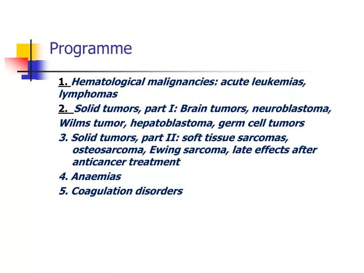
Programme. 1. Hematological malignancies: acute leukemias, lymphomas 2. Solid tumors , part I: Brain tumors , neuroblastoma , Wilms tumor , hepatoblastoma , germ cell tumors

E N D
Programme 1. Hematological malignancies: acute leukemias, lymphomas 2. Solid tumors, part I: Brain tumors, neuroblastoma, Wilmstumor, hepatoblastoma, germ cell tumors 3. Solid tumors, part II: soft tissue sarcomas, osteosarcoma, Ewing sarcoma, late effects after anticancer treatment 4. Anaemias 5. Coagulation disorders
Pediatric oncology and hematology Hematological malignancies: • Acute lymphoblastic leukemia • Acute non-lymphoblastic (myeloblastic) leukemia • Non-Hodgkin lymphoma • Hodgkin lymphoma
Uncontrolled proliferation of immature blood cells with a different immunological subtypes which is lethal within 1 –6 months without treatment The disorder starts in the bone marrow, where normal blood cells are replaced by leukemic cells Morphological (FAB), immunological, cytogenetic, biochemical, and molecular genetic factors characterize the subtypes with various response to treatment
Incidence Most frequent neoplasm in children (28 – 33%) 45/ 1million children under the age of 16 years Incidence peak at 2 – 5 years 75-80%- acute lymphoblastic leukemia -ALL 15-20% - acute myelogenous (non-lymphoblastic) leukemia AML/ ANLL <5% - undifferentiated acute leukemia and chronic myelogenous leukemia -CML
Ethiology Unknown Higher risk in congenital disorders: -trisomy 21 (14 times higher) and other trisomies -Turner syndrome -Klinefelter syndrome -monosomy 7 -neurofibromatosis type 1 -Fanconi anemia (high fragility of chromosomes) -Bloom syndrome, Kostmann S., Shwachman-Diamond S., -ataxia- teleangiectasia -congenital agammaglobulinemia -Wiskott- Aldrich S.
Ionizing radiation (atomic bomb developed high incidence of leukemia) Chemical and drugs: -benzene -chloramphenicol -alkylating agents Infection (viral –HTLV, EBV, HIV) Immunodeficiency: agamma/hypogammaglobulinemia, Wiskott-Aldrich S, HIV infection
Acute lymphoblastic leukemia 80% of leukemias Girl – to- boy ratio is 1: 1.2 Peak incidence 2 – 5 years Incidence in white children is twice as high as in nonwhite children
Clinical manifestation General aspects: - history and symptomsreflect: the degree of bonemarrowinfiltration by leukemiccellsand the extramedullaryinvolvement of the disease - the duration of symptomsisdays to severalweeks, occasionally – severalmonths - often: low –gradefever, signs of infection, fatigue, bleeding, pallor
The symptoms depend on the degree of cytopenia: - anemia: pallor, fatigue, tachycardia, dyspnea, occasionally- cardiovascular decompensation - leukopenia:infections, temperature elevation - thrombocytopenia: petechiae, mucosal bleeding, epistaxes, prolonged menstrual bleeding
Specific signs and symptoms Eye: bleeding, infiltration of local vessels,
CNS: at time of diagnosis less than 5% have CNS leukemia with meningeal signs (morning headache, vomiting, papilla edema, focal neurological signs)
Ear, nose, throat: -lymph nodes infiltration (isolated or multiple) -Mikulicz syndrome (infiltration of salivary glands and/or tear glands) Laboratory: Lk 3,91 Er 2,91 Hb 9,2 Ht 25,1 Blood smear: neutr-9% ly- 45% bl-46% LDH 976
Skin: maculopapular skin infiltration, often of deep red color (infants)
Cardiac involvement: -leukemic infiltration or hemorrhage -occasionally cardiac tamponade due to pericardial infiltration -tachycardia, low blood pressure or other signs of cardiac insufficiency Mediastinum: -enlargement due to leukemic infiltration by lymph nodes and /or thymus (observed in T-cell leukemia) Pleura/and pericardium: effusion Kidney enlargement
Lymphadenopathy Gastrointestinal involvement: -hepato- and/or splenomegaly
Testicular involvement: enlargement of one or both testes without pain , hard consistency Penis: priapism is occasionally associated with elevated WBC Bone and joint involvement: -bone pain initially present in 25 % to 50% of patients ! -bone or joint pain, sometimes with swelling and tenderness due to leukemic infiltration of the periosteum. Differential diagnosis: rheumatic fever, rheumatoid arthritis -radiological changes: diffuse demineralization, osteolysis,
Laboratory findings Red cells: -hemoglobin – normal/ moderate /markedly low -low number of reticulocytes White blood cell : - normal/ low/ high -in children with high WBC- leukemic blast cells present Platelets: -usually low
Coagulopathy: -in children with hyperleukocytosis -more common in AML -low levels of prothrombin, fibrinogen, factors V, IX, and X may be present Chemistry: -the serum uric acid is often high initially - the serum potassium level may be high (cell lysis) -serum hypocalcemia or hypercalcemia (in marked leukemic bone infiltration) abnormal liver function > increased level of transaminases
Bone marrow analysis: >25% blasts-characterize the blast cells -determine the degree of reduction of normal hematopoiesis -morphological, immunological, biochemical, and cytogenetic analyses Differential diagnosis: aplastic anemia, myelodysplastic syndrome, neoplastic infiltrations (neuroblastoma, NHL)
Leukemic cell characterization and classification: Morphology: FAB classification: ALL - L1, ALL-L2, ALL-L3 AML M0 – M7 chemistry: ALL: + periodic acid Schiff(PAS) AML: + Sudan black, + peroxidase
Immunological characterization: -monoclonal antibodies to leukemia-associated antigens differentiate between types of leukemic cells: * lympoid stem cells: CD19, HLA-DR, CD 24 (+/-) * early pre-B cells: CD19, HLA-DR, CD24 * pre-B cells: CD19, HLA-DR, CD24, CD10, CD20(+/-) * B-precursors cell: CD19, HLA-DR, CD24, CD10, CD20 * T-cell lineage: CD7, CD2, CD1, CD4, CD8,CD3
Cytogeneticcharacterization: - in 85% of childrenabnormalkaryotypeinthemalignant clone *t(9;22) (BCR-ABL) –unfavorableprognosis *t (4;11) ininfants , poorprognosis -ploidy and structure of chromosomes (rearrangements) -hypoploidy- poorprognosis -DNA index (DI)
Prognostic factors Favorable: WBC <10x10 9/l Age 2-7 Female Response on steroid (+) Pre-B-ALL Hyperploid FAB L1 ↑LDH moderate Unfavorable: WBC >50 x 10 9/L Age < 2 and >10 Male Response on treatment (-) Hypoploid, t(9;22)/t(9;11) FAB L2/L3 ↑↑LDH high visceromegaly
Differential diagnosis Leukemic reaction in bacterial infection, acute hemolysis,tuberculosis, sarcoidosis, histoplasmosis Lymphocytosis: pertussis Infectious mononucleosis Aplastic anemia Idiopathic thrombocytopenia Bone marrow infiltration by a solid tumor (NBL,NHL, RMS) Rheumatoid arthritis, rheumatoid fever
Therapy In experienced center Subdivided into: -remission induction -consolidation with CNS prophylaxis -maintenance phase Prognosis Rate of first remission in ALL: more than 90% 80% of children survive without relapse
Response on treatment • Reaction on steroids (7.day) • Reaction on chemotherapy (15. and 33. day) • Minimal residual disease - MRD (-)
Acute myelogenous leukemia Heterogeneous group of malignant hematological precursor cells of the myeloid, monocytic, erythroid or megakaryocytic cell lineage Epidemiology: 15-20% of all leukemias in children Frequency remains stable throughout childhood with slight increase during adolescence No difference in incidence between boys and girls
FAB classification M0: immature myeloblastic leukemia M1: myeloblastic leukemia M2: myeloblastic leukemia with signs of maturation M3: promyelocytic leukemia M4: myelomonocytic leukemia M5: monocytic leukemia M6: erythroleukemia M7: megakaryocytic leukemia
Clinical presentation Bleeding: thrombocytopenia + coagulopathy (DIC) Leukostasis in the lungs or CNS Tumor lysis syndrome Granulocyticsarcoma (chloroma) Infection (fungal, opportunistic)
Therapy Induction/ consolidation/ intensification/ maintenance - in AML3 + ATRA Allogeneic/ autologous stem cell transplantation Prognosis 5+year survival rate 50-60 %
Neoplasia of the lymphatic system and its precursor cells with genetically disturbed regulation, differentiation and apoptosis If marked bone marrow involvement is present the clinical condition is equal of leukemia Incidence 5 –7 % of all neoplasias in childhood Peak incidence between 5 and 15 years Ratio of boys to girls 2:1 Burkitt lymphoma (BL): endemic form in Africa 10:100,000 children and sporadic form in Europe and USA
Etiology, pathogenesis and molecular genetics Often chromosomal alterations are detecable: in B-cell NHL translocation of chromosome 14 - t(18;14) Predisposing factors for NHL: Acquired immunodeficiency: autoimmune disorders, HIV infection EBV infection Congenital B-cell defect, congenital T-cell defect with thymus hyperplasia Bloom syndrome, Chedak-Higashi syndrome, SCID, ataxia teleangiectasia, Wiskott-Aldrich syndrome Exposure to irradiation Drug induced, after immunosuppressive treatment
Burkitt lymphomaBurkit-like lymphoma About 50% of NHL Localization: abdomen, lymphatic tissue of adenoids and tonsils 80% with translocation t(8;14) or t(8;2) and t(22;8) with c-MYC on chromosome 8q24 which stimulates proliferation 40% with a p53 mutation
Large B-cell lymphoma 7-8% of NHL Localization: abdomen, peripherallymphnodes, skin, bone Lymphoblasticlymphoma 30% of NHL Usuallymediastinallocalization AnaplasticLargeCellLymphoma 7-8% of NHL
Clinical manifestations Duration of symptoms: usually a few days to weeks Non-specific symptoms: fatigue, nausea, anorexia, loss of weigth and/or fever In relation to localisation of NHL: Abdomen: especially the ileocecal region, mesentery, retroperitoneum, ovaries > painfull, spasms, vomiting Obstipation, intussusception Apendicitis-like Ileus, ascites
Mediastinum: Mostly anterior or middle part of mediastinum > cough, stridor, dyspnea, wheezing Edema of the neck and face with marked dyspnea may indicate SVCS Pain of the back or abdomen Pleural effusion
Involvement of adenoid and tonsils, nasopharyngeal lymph nodes, parotid gland swelling
Peripheral lymph nodes: Mostly cervical, supraclavicular and inguinal Lymph nodes are firm, not usually tender, but involving multiple lymph nodes that usually occur unilaterally Other locations: CNS, cranial and peripheral nerves, skin, muscles, bone, thorax, gonads, parotid gland, epidural region→ spinal cord compression
Differential diagnosis Lymph node enlargement in infectious diseases Autoimmune lymphoproliferative syndrome Hodgkin Lymphoma metastatic disease of sarcomas or neuroblastomas ALL: if more than 25% blasts = ALL, if less= NHL IV stage
Diagnosis:-histology-stage Histological (lymph nodes, peripheral blood, bone marrow or fluid resulting from pleural effusion or ascites) In abdominal stage: laparotomy In SVCS- emergency situation, noninvasive biopsy or pretreatment with chemotherapy or/and radiotherapy Morphological, immunophenotypical and molecular /cytogenetic analyses Serum lactate dehydrogenase (LDH) Serum uric acid Bone marrow aspiration CSF analysis
Radiological diagnosis Ultrasound Conventional X-ray CT of the thoracic, abdomen and skeletal disease MRI for CNS PET (positron –emmision tomography) Bone scan
Staging ( Murphy/St.Jude) • I- a single tumor (extranodal) or single anatomical area (nodal), excluding mediastinum or abdomen • II- a single tumor (extranodal) with regional involvement On same side of diaphragm a/ two or more nodal areas b/ two single (extranodal) tumors with or without regional node involvement A primary gastrointestinal tract tumor (usually ileocecal) with or without associated mesenteric node involvement; gross complete resection
III- On both sides of the diaphragm: a/two single tumors (extranodal) b/two or more nodal areas ALL primary intrathoracic tumors (mediastinal, pleural, thymic) All extensive primary intra-abdominal disease, unresectable All primary paraspinal or epidural tumors regardless of other sites • IV- Any of the above with initial CNS or bone marrow involvement (less than 25%)