Download
1 / 25
280 likes | 978 Views
Management of Acute Bleeding from a Peptic Ulcer. N Engl J Med 2008;359:928-37. Epidemiology. The vast majority of acute episodes of UGI bleeding (80 to 90%) have nonvariceal causes, with peptic ulcer accounting for the majority of lesions.

E N D
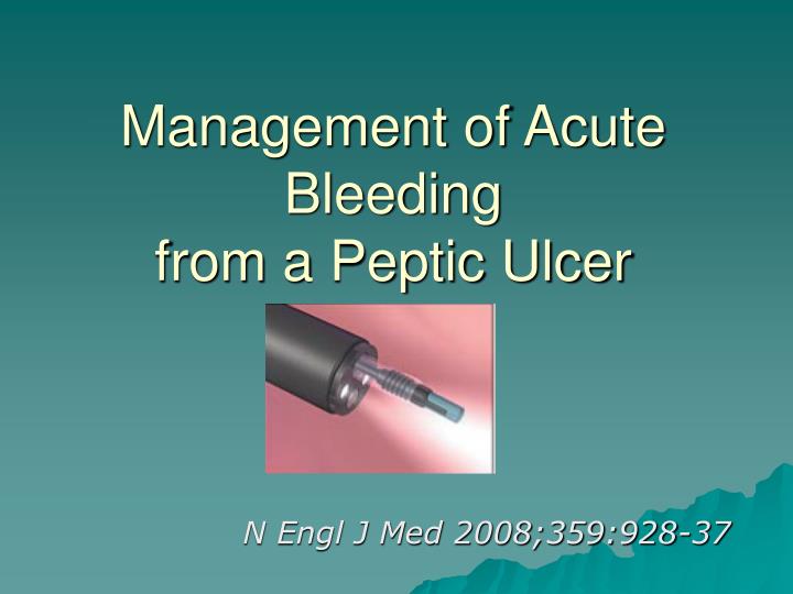
Management of Acute Bleedingfrom a Peptic Ulcer N Engl J Med 2008;359:928-37
Epidemiology • The vast majority of acute episodes of UGI bleeding (80 to 90%) have nonvariceal causes, with peptic ulcer accounting for the majority of lesions. • The annual incidence of bleeding from a peptic ulcer may be decreasing worldwide ( the incidence is 60 per 100,000 population) • an increasing proportion of episodes related to the use of aspirin and NSAID • Peptic ulcer bleeding is seen predominantly among the elderly • 68% > 60Y/O and 27% >80Y/O • Mortality associated with peptic ulcer bleeding remains high at 5 to 10%
Management of Acute Bleeding from a Peptic Ulcer, According to Clinical Status and Endoscopic Findings
The first priority in treatment is correctingfluid losses and restoring hemodynamic stability.
Initial Management • Assess hemodynamic status • Tachycardia (pulse, ≥100 beats per minute) • Hypotension (systolic blood pressure, <100 mm Hg), • postural changes (an increase in the pulse of ≥20 beats per minute or a drop in systolic blood pressure of ≥20 mm Hg on standing) • Mucous membranes, neck veins, urine output • Obtain CBC, electrolytes, BUN/Cr, PT INR/ APTT, blood type, and cross-match
Initial Management • Initiate resuscitation with crystalloid intravenous fluids with the use of large-bore IV-access catheters • two peripheral catheters of 16 to 18 gauge • a central catheter if peripheral access is not available • PRBC • If tachycardia or hypotension is present • If the hemoglobin level is less than 10 g per deciliter. • Oxygen • correction of coagulopathy
The insertion of a NG tube • The presence of red blood in the NG aspirate is an adverse prognostic sign • 15% of patients without bloody or coffee-ground material in NG aspirates are found to have high-risk lesions on endoscopy • The use of a large-bore OG tube with gastric lavage • improve visualization of the gastric fundus on endoscopy • not improve the outcome. • No role for occult-blood testing of aspirate
Initial Management • Consider giving a single 250-mg IV dose of erythromycin 30 to 60 minutes before endoscopy • promote gastric motility and substantially improve visualization of the gastric mucosa on initial endoscopy. • not improve the diagnostic yield of endoscopy substantially or to improve the outcome • Consider initiating treatment with an IV PPI (80-mg bolus dose plus continuous infusion at 8 mg/hr) while awaiting early endoscopy • down-staging of endoscopic lesions • not have an effect on outcomes • The cost- effectiveness remains controversial • No role for H2 blocker
Risk-Stratification Tools for Upper Gastrointestinal Hemorrhage • The Rockall score : • Used clinical and endoscopic criteria • The scale ranges from 0 to 11 points, with higher scores indicating higher risk. Blatchford scores from 0 to 23, with higher scores indicating higher risk
Early endoscopy • The cornerstone of treatment • performed within 24 hours • Improve certain outcomes • the number of units of blood transfused • the length of the hospital stay • Determine the cause of bleeding, ascertain prognosis, and administer endoscopic therapy. • Treatment recommendations have focused on the first 72 hours after presentation and endoscopic evaluation and therapy, since this is the period when the risk of rebleeding is greatest (90 %)
Forrest classification Forrest grade IA Forrest grade IB Forrest grade IIA
High-risk — Forrest grade IA, IB, or IIA • Perform endoscopic hemostasis • contact thermal therapy • mechanical therapy ( hemoclips ) • epinephrine injection, followed by contact thermal therapy or by injection of a second injectable agent. • Epinephrine injection as definitive hemostasis therapy is not recommended • The endoscopist should use the most familiar hemostasis technique
High-risk — Forrest grade IA, IB, or IIA • Admit the patient to a monitored bed or ICU setting(for the first 24 hours of what is usually at least a 3-day hospital stay) • Treat with an IV PPI (80-mg bolus dose plus continuous infusion at 8 mg per hour) for 72 hours after endoscopic hemostasis • No role for H2 blocker, somatostatin, or octreotide. • Initiate oral intake of clear liquids 6 hours after endoscopy in patients with hemodynamic stability • Transition to oral PPI after completion of IV therapy. • Perform testing for Helicobacter pylori; initiate treatment if the result is positive.
Effect of Proton-Pump Inhibition in Peptic-Ulcer Bleeding • Gastric acid impairs clot formation, promotes platelet disaggregation, and favors fibrinolysis • Inhibiting gastric acid and raising the intragastric pH to 6 or more and maintaining it at that level may promote clot stability, thus decreasing the likelihood of rebleeding. • Although data from clinical trials support the use of a bolus followed by a continuous infusion of proton-pump inhibitors, recent studies from North America show that even a high-dose, continuous infusion of proton-pump inhibitors may not sustain an intragastric pH of 6 or more. • The reduction in mortality appears to occur only among patients with high-risk stigmata who have first undergone endoscopic therapy, a finding that supports the use of medical therapy as an adjunct to but not a replacement for endoscopic hemostasis.. • Intravenous bolus loading followed by continuous infusion of proton-pump inhibitors is more effective than bolus dosing alone in decreasing the rates of rebleeding and the need for surgery
High-dose oral PPI • The use of high-dose oral PPI in peptic-ulcer bleeding has been shown in Asian populations reductions • the risk of rebleeding • the need for surgery • the risk of death • These results may not be completely generalizable to North American or European populations
Second-look endoscopy • Planned, second-look endoscopy that is performed within 24 hours after initial endoscopic therapy has not been recommended. • It provided only a limited reduction in the rate of rebleeding. (Two meta-analyses) • It may not be cost-effective when medical therapy leading to profound acid suppression is used. • Repeat endoscopy may be considered on a case-by-case • if there are clinical signs of recurrent bleeding • if there is uncertainty regarding the effectiveness of hemostasis during the initial treatment.
Forrest classification Forrest grade IIB
High-risk — Forrest grade IIB • Consider endoscopic removal of adherent clot, followed by endoscopic hemostasis if underlying active bleeding or nonbleeding visible vessel is present. • Admit the patient to a monitored bed or ICU setting. • Treat with IV PPI (80-mg bolus dose plus continuous infusion at 8 mg per hour) for 72 hours after endoscopy, regardless of whether endoscopic hemostasis was performed • No role for H2 blocker, somatostatin, or octreotide • Initiate oral intake of clear liquids 6 hours after endoscopy in patients with hemodynamic stability • Transition to an oral PPI after completion of IV therapy. • Perform testing for H. pylori; initiate treatment if the result is positive.
Forrest classification Forrest grade IIC Forrest grade III
Low-risk — Forrest grade IIC or III • Do not perform endoscopic hemostasis. • Consider early hospital discharge after endoscopy if the patient has an otherwise low clinical risk and safe home environment. • Treat with an oral PPI. • Initiate oral intake with a regular diet 6 hours after endoscopy in patients with hemodynamic stability. • Perform testing for H. pylori; initiate treatment if the result is positive.
Predictors of failure of endoscopic treatment • History of peptic ulcer disease • Previous ulcer bleeding • The presence of shock at presentation • Active bleeding during endoscopy • Large ulcers (>2 cm in diameter) • Large bleeding vessel (≥2 mm in diameter) • Ulcers located on the lesser curve of the stomach or on the posterior or superior duodenal bulb
After endoscopy • If there is clinical evidence of ulcer rebleeding, repeat endoscopy with an attempt at endoscopic hemostasis,obtain surgical or interventional radiologic consultation for selected patients. • For selected patients, discuss the need for ongoing use of NSAIDs, antiplatelet agents, and concomitant therapy with a gastroprotective agent.