Download
1 / 41
420 likes | 799 Views
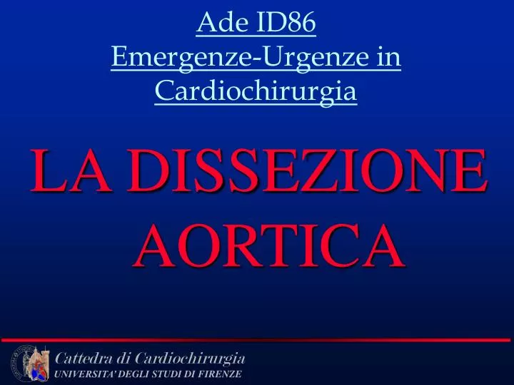
Ade ID86 Emergenze-Urgenze in Cardiochirurgia. LA DISSEZIONE AORTICA. Dissezione Aortica. red blood clot. true lumen. false lumen. Scissione longitudinale della media aortica da parte di una colonna di sangue

E N D
Ade ID86Emergenze-Urgenze in Cardiochirurgia LA DISSEZIONE AORTICA
Dissezione Aortica red blood clot true lumen false lumen
Scissione longitudinale della media aortica da parte di una colonna di sangue • La scissione della media occupa circa metà della circonferenza dell’aorta e può estendersi per tutta la lunghezza del vaso • Lo scollamento creato dal sangue crea un secondo lume o falso lume
Dissecting aortic aneurysm (aortic dissection). This gross picture highlights a 6cm wide aortic intimal and partial medial tear just above the aortic valve. Most dissections start with a intial tear in the first 10cm of the aorta. Through this tear blood enters, and dissects down the laminar plain of the media. It often will re-enter the vessel lumen in the abdominal aorta or even lower (illiac or femoral arteries).
Dissecting aortic aneurysm. This is a segment of the abdominal aorta showing the two seperated walls of the dissecting aneurysm (the blood has been removed). The media is seperated easily, like the pages of a book. Inner 1/3 of media Outer 1/3 of media with adventitia
Epidemiologia • Incidenza: 1caso/80.000abitanti • 350 nuovi casi/anno • Età: 40-70 anni • M/F: 3/1
CLASSIFICAZIONE Classificazione di De Bakey • Tipo I: dissecazione dal tratto ascendente al discendente • Tipo II: dissecazione limitata all’arco a al tratto ascendente, senza coinvolgimento del tratto discendente Tipo III: dissecazione della sola aorta discendente
CLASSIFICAZIONE Classificazione di Stanford Tipo A: interessamento aorta ascendente Tipo B:interessamento arco aortico e/o aorta discendente, non l’ascendente
Fattori predisponenti • Ipertensione (75-90% dei casi) • S. di Marfan e Collagenopatie • Malformazioni aortiche congenite • Valvulopatie aortiche acquisite • Gravidanza (rara) • Forme iatrogene (cateterismi) • Abuso di Cocaina
Quadri clinici e sintomatologia • Lacerazione e sede della lacerazione • Progressione dello scollamento con coinvolgimento aa. collaterali • Sindrome da compressione • Sindrome da rottura
Sintomi • DOLORE (Migrante) • 50% STENOCARDICO 30% ADDOMINALE • 10% DORSALE 6% LOMBARE
The pain of dissection is usually sudden, worst at onset, severe, and not previously experienced. Adjectives such as “ripping” and “tearing” often are used by the patient
SINTOMI LEGATI ALLA PROGRESSIONE • Right carotid compression from dissected blood • Blood dissected upward from aortic tear • Can dissect upward toward carotids or downward toward coronaries • Therefore, symptoms may reflect those of a stroke or heart attack
SINTOMI LEGATI ALLA PROGRESSIONE • SINDROMI ISCHEMICHE DELL’ARCO • -disturbo della coscienza • -lipotimia • -mono- emiparesi • -plegie • -scomparsa polsi periferici arti sup. • -asimmetrie di pressione dx-sx • OCCLUSIONE AA.SPINALI • -parestesie • -paraplegie • -paraparesi. • OCCLUSIONE AA.RENALI • -anuria • -ematuria • -infarto renale • -ipertensione nefrovascolare • -dolore (colica renale) • OCCLUSIONE AA. MESENTERICHE • -dolori addominali • -infarto intestinale • OCCLUSIONE AA. ILIACHE • -simula ischemia acuta dell’arto • -sciatalgia
SINDROME DA COMPRESSIONE • S. Mediastinica per compressione n. laringeo ricorrente • S. di Horner per compressione del ganglio stellato • S. da compressione esofagea con disfagia
Blood dissected proximally through the media • Hemopericardium resulted • Cardiac tamponade may be present due to the extreme hemorrhage SINDROME DA ROTTURA • Pericardio • Cavità Pleurica • Addome
PROGNOSI • Acute Type A dissection is a surgical disease • Previous natural history studios show 80-90% one month mortality • Two recent series have indicated a 60% one month mortality with non-operative medical therapy • Senza trattamento, Ao dissecazione ha un’alta mortalità • Il 35% dei pz non curati muore entro le prime 24 ore, il 50% entro 48 ore, il 70% dopo una settimana e l’80% entro 2 settimane. • Il tipo B ha prognosi migliore STORIA NATURALE DELLA DISSEZIONE SOPRAVVIVENZA TIPO B SOPRAVVIVENZA TIPO A
Iter diagnostico iniziale nel pz con sospetta dissezione aortica • Esami ematochimici(CPK,CK-MB, T(I)troponina, conta bianchi, D-dimero, emocromo, LDH…) • ECG • RX TORACE • ECO
ESAMI DIAGNOSTICI Laboratory data(1) 1. Decreases in the hemoglobin and hematocrit are ominous findings suggesting the dissection either is leaking or has ruptured. 2. BUN and creatinine are elevated if the dissection involves the renal arteries. 3. Hematuria, oliguria, and even anuria (<50 mL/d) may occur if the dissection involves the renal arteries. 4. CKMB and Troponin T may be elevated in acute thoracic aorta dissection
ESAMI DIAGNOSTICI Laboratory data(2) 1. In acute thoracic dissection, ECG can mimic the changes seen in acute cardiac ischemia. In the presence of chest pain, these signs can make distinguishing dissection from AMI very difficult. Keep this in mind when administering thrombolytics to patients with chest pain. STT depression and T wave inversion(red arrow )
1. Chest X-ray is used as routine screening 2. Contrast-enhanced CT can image arch and descending aorta 3. MRI if available is usually best for imaging ascending aorta 4. Transesophageal ultrasound, if available, especially for root and ascending aorta 5. Angiography is more invasive and has been replaced by many other imaging such CT, MTI Aortography sens-88% spec-94% CT sens-83% spec-100% ( noninvasive, need contrast, 3D capabilities) MRI sens-98% spec-98% (noninvasive, no contrast ) ECHO -TTE sens 59-85% spec-63-96% ECHO -TEE sens 98% spec 98% ( important disadvantage of TEE is its limited ability to visualize the distal ascending Ao and proximal arch because of interposition of the air filled trachea and main stem bronchus.) Diagnostica per immagini Selection of imaging diagnosis
left pleural effusion mediastinum widening Diagnostica per immagini(1) Chest X-ray 1. Mediastinum widening 2. Displacement of intima calcification 3. Displacement of endotrachea tube and NG tube 4. Left pleural effusion (signs of dissecting ruptire)
Diagnostica per Immagini (2) CT scan 1. Intimal flap 2. Displacement of intimal calcification 3. Differential contrast enhancement of true v.s. false lumen T Intimal flap F
Partition of a three-dimensional contrast-enhanced MRA shows intimal flap (arrows ) in the distal aortic arch and descending aorta. Diagnostica per Immagini(3) MRI 1. Intimal flap 2. Slow flow and clot in false lumen
Diagnostica per Immagini(4) Transesophgeal echocaediogram 1. Freely movable flap within the lumen of the vessel 2. Differential Doppler detection of true v.s. false lumen F T Freely movable flap within the aorta
A dissection flap can be seen spanning the aneurysmal ascending aorta. A defect is noted in the central part of the flap consistent with an entry point from the true into the false lumen. Colour flow Doppler demonstrates the communication between true and false lumens.
On 2-D imaging a dissection flap (arrows) is seen within the descending thoracic aorta. A PW Doppler cursor has been placed in each of the two lumens shown above. Flow is greater in the true lumen (left) than in the false lumen (right). Colour flow Doppler confirms the smaller, more posterior lumen, as the true lumen.
F Diagnostica per Immagini (5) Angiography 1. Intimal flap 2. True and false lumen (may be failure if the false channel is thrombosed) 3. Aortic regurgitation 4. Coronary artery T Oblique arteriogram of the thoracic aorta demonstrates the double-barrel aorta sign of aortic dissection. Both the true and false lumina are opacified
PROGNOSI STORIA NATURALE DELLA DISSEZIONE Senza trattamento, la dissecazione ha un’alta mortalità: • prime 24 ore35% • entro 48 ore50% • dopo una settimana 70% • entro 2 settimane80%. Il tipo B ha prognosi migliore
DIAGNOSI E TRATTAMENTO DELLA DISSEZIONE AORTICA STABILITÀ EMODINAMICA INSTABILITÀ EMODINAMICA Sala operatoria Diagnosi sospetta Anestesia generale + monitoraggio (TEE) Diagnosi certa Terapia Intensiva Negativo Positivo Conferma diagnosi (TEE) Diagnosi sospetta Diagnostica inadeguata Intervento chirurgico TAC Pos Neg RMN
TERAPIA (1) • Pz in ACR con PEA: RCP Considerare PERICARDIOCENTESI
TERAPIA (2) Dissezione aortica tipo A • Terapia Chirurgica URGENTE • Any dissection involving the ascending aorta • Symptomatic or complicated descending aortic dissections
TERAPIA (3) Dissezione aortica tipo B • Terapia Medica • Per ridurra P.A. e stress sulla parete arteriosa • B bloccanti • Vasodilatatori • Per il dolore morfina, NO FANS • Terapia Chirurgica • Symptomatic or complicated descending aortic dissections
TYPE A AORTIC DISSECTION tear location true lumen false lumen The ruler has been inserted into the tear in the intima that has given rise to the dissection of this vessel. This is a view of the intimal surface of the aorta TYPE A AORTIC DISSECTION clots in false lumen and aortic arch reentry TYPE A AORTIC DISSECTION clots in false lumen
A Cabrol interostial coronary graft or an ultra short (1.5-cm) graft-to-left-main extension occasionally is employed when low-lying ostia cannot be mobilized.