Download
1 / 59
600 likes | 1.44k Views
Abdominal Pain and Bowel Obstruction. Mike Goodwin CRASH Course October, 2010. Abdominal Pain - Approach. History Physical Labs Imaging Provisional Dx. History. PQRST AAA etx But don’t forget PSx Bowel/ Gyne / Urol ROS. Physical Exam. Complete

E N D
Abdominal Pain and Bowel Obstruction Mike Goodwin CRASH Course October, 2010
Abdominal Pain - Approach History Physical Labs Imaging Provisional Dx
History • PQRST AAA etx • But don’t forget • PSx • Bowel/Gyne/Urol ROS
Physical Exam • Complete • General appearance/vitals/H+N/Chest • Abdo: • Rigidity • Rebound • Guarding • IPPA • DRE / Pelvic / Groin / Flank-CVA
Labs • Everyone: • CBC, lytes BUN Cr • LFT, Bili, Amylase/Lipase, lactate • Urinalysis • Urine Preg
Imaging • AXR • 3-views • Free air • Distended bowel/air-fluid • Calcifications (panc or kidney/ureter) • US • If GS disease suspected • Lower abdo pain in female
Imaging • CT Abdo • Test of choice for most patients • Protocols to minimize contrast nephropathy
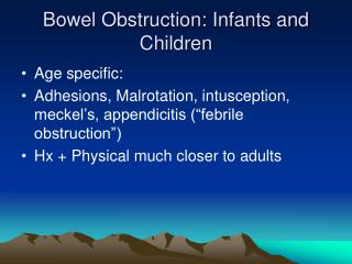
Bowel Obstruction: Overview • History • Etiology • Pathophysiology • Clinical presentation • Imaging • Management • Special considerations
Causes of Small Bowel Obstruction in Adults • Lesions Extrinsic to the Intestinal Wall • Lesions Intrinsic to the Intestinal Wall • Intraluminal/Obturator Obstruction
Lesions Extrinsic to the Intestinal Wall • Adhesions (usually postoperative) • Neoplastic • Carcinomatosis • Extraintestinal neoplasms • Hernia • External (e.g., inguinal, femoral, umbilical, or ventral hernias) • Internal (e.g., congenital defects such as paraduodenal, foramen of Winslow, and diaphragmatic hernias or postoperative secondary to mesenteric defects • Intra-abdominal abscess
Congenital Malrotation Duplications/cysts Inflammatory Crohn’s disease Infections Tuberculosis Actinomycosis Diverticulitis Neoplastic Primary neoplasms Metastatic neoplasms Traumatic Hematoma Ischemic stricture Miscellaneous Intussusception Endometriosis Radiation enteropathy/stricture Lesions Intrinsic to the Intestinal Wall
Intraluminal/Obturator Obstruction • Gallstone • Enterolith • Bezoar
Common causes of small bowel obstruction in industrialized countries.
Pathophysiology • Early: Increased motility & contractility • Bowel dilation, fluid/lytes accumulate in lumen and bowel wall • Third spacing, intravascular volume depletion
Bowel obstruction Increased intraluminal pressure Decreased mucosal blood flow Progressive Ischemia Perforation & Peritonitis
Clinical Diagnosis • History • Colicky abdominal pain • Nausea / vomiting • Abdominal distension • Failure to pass flatus / feces
Physical Examination • Vitals: Tachycardia, hypotension • Abdomen: • Distension • Surgical scars • Bowel sounds, increased or decreased • Localized tenderness / rebound / guarding suggests strangulation • Hernia exam (ventral, groin, etc) • Rectal exam: • Rectal masses • Blood – suggesting ischemia, malignancy
Radiology • Plain Abdo X-Rays • Confirm Diagnosis • Localize obstruction to small bowel or colon • Evidence of complete or incomplete
Plain X-ray Features • Dilated Small Bowel (>3 cm) • Multiple air-fluid levels • Colonic gas pattern • Normal / Dilated (Ileus or partial obstruction) • Absence of gas c/w complete obstruction • *Thickened bowel wall • *Pneumatosis intestinalis *Suggests ischemia/strangulation
Plain X-rays • Lappas et al 2001 • Review of 12 AXR findings with SBO • Findings: • Combination of • Air-fluid levels of different heights in the same bowel loop • Mean air-fluid level diameter of 2.5 cm or greater • Most predictive of a high-grade partial or complete SBO
AXR Disadvantages • 20-30% false negative rate • Does not localize site of obstruction • Does not establish etiology of obstruction
CT Scan • 95% sensitive • 96% specific • 95% accurate in determining the presence of complete or high-grade SBO • Shows site and cause of obstruction in 95% of instances • Less accurate for partial SBO (50% some studies)
CT for SBO • CT performed with IV and PO contrast • High-grade SBO seen even with no contrast • Lesser grades of obstruction seen with PO contrast • IV contrast for assessment of bowel wall for signs of edema or ischemia.
When to Order CT? • Clinical presentation or abdominal films nondiagnostic • Hx of abdominal malignancy • Immediate postsurgical patients • Patients who have no history of abdominal surgery
Barium / Contrast Studies • History of recurring obstruction • Low-grade mechanical obstruction • Defines the obstructed segment and degree of obstruction
Gastrograffin Swallow in Adhesive SBO, Cochrane Review, 2004 • Diagnostic • Gastrofraffin seen in the cecum on AXR within 24 hours predicts resolution • Sensitivity of 0.96, specificity of 0.96 • Therapeutic • Hospital length of stay 2-3 days shorter in non-operative patients • Studies prospective, non-blinded
Simple Versus Strangulating Obstruction • Classic signs: • Fever • WBC inc • Constant Abdo pain • But no parameters reliably detect strang. • CT findings detect late ischemic changes
Treatment – Nonoperative • Fluid resuscitation • IV resuscitation with isotonic saline • Electrolyte replacement • Monitor urine output • Tube decompression • Empties stomach • Reduces aspiration risk • No benefit to long intestinal tubes • In partial obstruction: 60-85% success rate
Treatment - Operative • Complete obstruction • Generally mandates operation • Some have argued for nonoperative approach in selected patients • 12-24hr delay of surgery is safe • >24hr delay is unsafe
Operative Technique • Dependent on underlying problem • Adhesive band: Lysis of adhesions • Incarcerated hernia: manual reduction and closure of defect • *Presence of hernia with SBO mandates OR • Malignant tumors: Difficult challenge • Diverting stoma • Resection / anastamosis • Enteroenterostomy
Intestinal Viability at Surgery • Release obstructed segment • Place in warm sponge x 15-20 minutes • If normal colour and peristalsis: return to abd • Doppler probe adds little to clinical judgment (Bulkley, 1981) • Fluorescein may be useful in difficult cases • “Second look” in 24 hrs if questionable viability or if clinically deteriorates post-op
Laparoscopy in Acute SBO? • Criteria: • Mild distension • Proximal obstruction • Partial obstruction • Anticipated single-band obstruction • No matted adhesions / carcinomatosis
Special Considerations: Recurrent Adhesions • Multiple agents have been tried, none successful • Hyaluronate-based membrane shown to reduce severity of adhesion formation (Becker, 1996; Vrigland, 2002) • No studies yet to show reduction in obstruction
Special Considerations: Recurrent Adhesions • So far, best evidence to prevent adhesions is good surgical technique: • Gentle handling of bowel • Avoid unnecessary dissection • Exclusion of foreign material from peritoneum • Adequate irrigation / removal of debris • Place omentum around site of surgery
Special Considerations: Acute Post-op Obstruction • Obstructive symptoms after an initial return of bowel function and resumption of oral intake • Technical complication versus adhesions • CT scan useful to evaluate for complications: • Anastamotic leak • Narrow anastomosis • Internal hernia • Obstruction at stoma • Early reoperation may be indicated
Acute Adhesive Postoperative Obstruction • Difficult to distinguish from ileus • Incidence 0.7% • Highest incidence on small intestine (3% – 10%) • Present as early as POD 4 • Usually partial SBO • CT preferred modality
Acute Postoperative Obstruction (Adhesive) • 80% spontaneous resolution of symptoms • 4% of patients required more than 2 weeks of treatment • SBO after laparoscopy: suspect herniaat trocar site
Surgery for Malignant Bowel Obstruction in Advanced Gynaecological and Gastrointestinal Cancer • Cochrane Review:2004 • Role of surgery controversial • No firm conclusions from many retrospective case series • Control of symptoms varies from 42% to over 80 • Rates of re-obstruction, from 10-50%, though time to re-obstruction was often not included • Continues to be a challenging problem
Steroids in Advanced Gyne/GI Cancer With SBO • Cochrane Review of prospective data (89 patients) • Trend, not statistically significant, for resolution of bowel obstruction using corticosteroids • No statistically significant difference in mortality • NNT 6 • Morbidity associated with steroids appears low